. 2011 Aug 14;18(9):1020-7.
doi: 10.1038/nsmb.2104.

A universal pathway for kinesin stepping

Affiliations

A universal pathway for kinesin stepping

Bason E Clancy et al. Nat Struct Mol Biol. .

Abstract

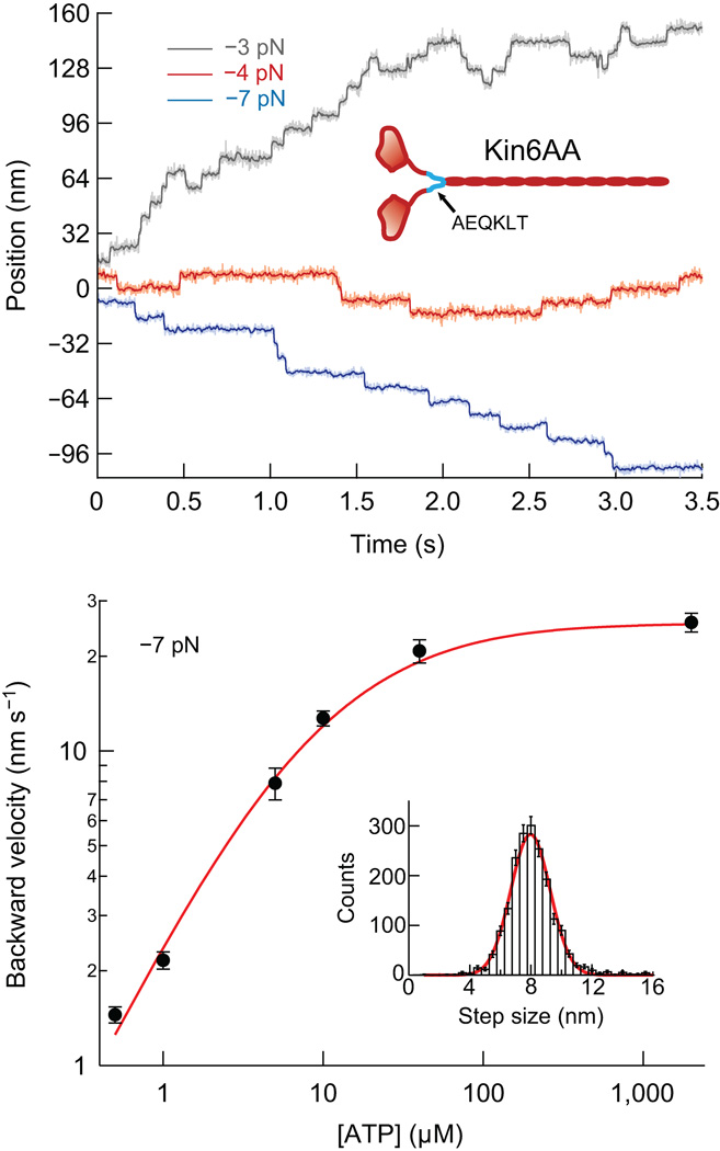
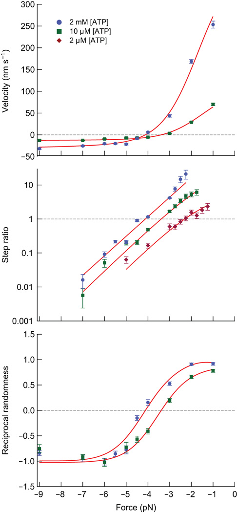
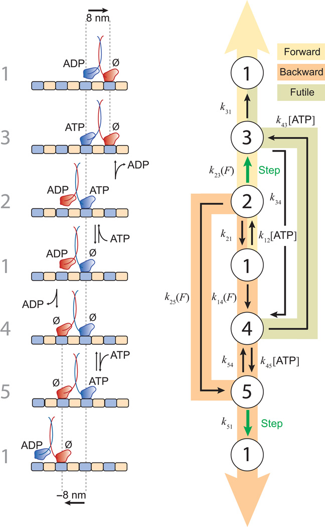
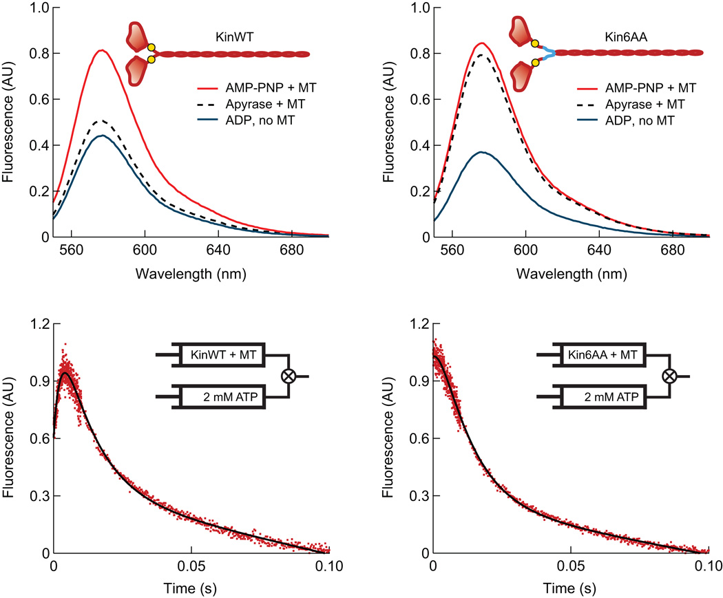
Kinesin-1 is an ATP-driven, processive motor that transports cargo along microtubules in a tightly regulated stepping cycle. Efficient gating mechanisms ensure that the sequence of kinetic events proceeds in the proper order, generating a large number of successive reaction cycles. To study gating, we created two mutant constructs with extended neck-linkers and measured their properties using single-molecule optical trapping and ensemble fluorescence techniques. Owing to a reduction in the inter-head tension, the constructs access an otherwise rarely populated conformational state in which both motor heads remain bound to the microtubule. ATP-dependent, processive backstepping and futile hydrolysis were observed under moderate hindering loads. On the basis of measurements, we formulated a comprehensive model for kinesin motion that incorporates reaction pathways for both forward and backward stepping. In addition to inter-head tension, we found that neck-linker orientation is also responsible for ensuring gating in kinesin.


Figures

Figure 1
Figure 1
Single-molecule records and backstepping velocity for Kin6AA. (a) Representative traces obtained under constant force at loads of −3 pN (gray), −4 pN (red), and −7 pN (blue), at 2 mM ATP. Light traces are unfiltered, darker traces are median-filtered. The records show clear 8-nm forward and backward steps. (b) Double-log plot of backstepping velocity vs. ATP concentration under −7-pN load (black dots; mean ± s.e.m.; N = 21−62). The solid line (red) shows the global fit to all single-molecule data for the model described in the text. Inset, histogram of the backward step size at a −7-pN load for all ATP concentrations (8.12 ± 1.72 nm, mean ± s.d., N = 1,906) and Gaussian fit (red line), centered at 7.97 nm.
Figure 2
Figure 2
Bi-directionality of Kin6AA as a function of load and ATP concentration. Solid lines (red) show the global fits to all single-molecule data for the model described in the text. (a) Velocity, v (mean ± s.e.m.) vs. force for 2 mM ATP (blue circles; N = 25−164) and 10 µM ATP (green squares; N = 18−74). Stall occurs where the fit data cross the horizontal dashed line (grey) at v = 0. (b) Step ratio, SR (mean ± s.e.m.; ratio of number of forward to backward steps) vs. force at 2 mM ATP (blue circles; N = 264−3,331), 10 µM ATP (green squares; N = 235−1,412), and 2 µM ATP (purple diamonds; N = 90−368). Stall occurs where the fit data cross the horizontal dashed line (grey) at SR = 1. (c) Reciprocal randomness, r−1, (mean ± s.e.m.), color-coded as in (a). Note that the fit data cross r−1 = 0 at the stall forces for the data in (a).
Figure 3
Figure 3
Model for stepping by kinesin dimers, showing forward stepping, backward stepping, and futile hydrolysis pathways. See text. (a) Left, the numbers assigned to each of the five states. The molecular configurations of the kinesin dimer on the microtubule thought to correspond to each of the states are illustrated, along with any nucleotides bound. No particular docking state of the neck-linker is implied in this diagram. Kinesin heads are color-coded (red, blue). Starting from State {1} (middle row), forward steps are accomplished by ascending the diagram; backward steps by descending. (b) Reaction diagram for the model, displaying the transition rates among states. Load- and ATP-dependent transitions are indicated. Three main pathways are shaded: forward stepping (yellow), backward stepping (orange), and futile hydrolysis (light green). Largely irreversible transitions between states that produce ±8-nm displacements are shown (green arrows). In this minimal model, fast transitions occurring in rapid succession were combined to generate composite states in several instances. Note: The transition from State {4} to State {3} in the futile hydrolysis pathway involves ATP binding to the rear head, but, unlike the stepping pathways, heads do not swap positions and no step is taken.
Figure 4
Figure 4
Fluorescence data for Kin6AA and KinWT with TMR probes attached to both neck-linkers. (a, b) Steady-state TMR fluorescence emission spectra for KinWT (a) and Kin6AA (b), which monitors neck-linker separation under the following conditions: microtubules plus 2 mM AMP–PNP (red), microtubules plus apyrase to remove nucleotides (black, dashed), and 2 mM ADP without microtubules (blue). The large signal increase in the absence of nucleotide (apyrase present) for Kin6AA is consistent with neck-linker separation. In the inset cartoons, approximate locations of the TMR probes (at position 333) are indicated (yellow circles), as well as the neck-linker inserts (blue lines). (c, d) Pre-steady-state TMR kinetic records for KinWT and Kin6AA. TMR-labeled motors complexed to a five-fold excess of microtubules and treated with apyrase were mixed with 2 mM ATP. The initial increase in fluorescence seen for KinWT is absent for Kin6AA, indicating that prior to mixing with ATP, both heads of Kin6AA are bound to the microtubule, and consequently, the neck-linkers of this mutant are separated.
Figure 5
Figure 5
Binding of 2′dmT to Kin6AA. (a) A complex of Kin6AA and microtubules was pre-formed and mixed in a stopped-flow apparatus with 2′dmT (Supplementary Methods). The resulting fluorescence signal (red) consisted of three sequential phases: a first phase of increasing fluorescence, a lag phase, and a second phase of increasing fluorescence. Fitting this signal required three exponential terms (black curve). Two terms, corresponding to the phases of increasing fluorescence, were associated with rate constants of 81.5 ± 21.0 s−1 and 3.0 ± 0.1 s−1. The third term was associated with a low-amplitude, decreasing phase, consistent with a lag, and a rate constant of 55.6 ± 24.8 s−1. The same experiment in the absence of microtubules (grey) produced a fluorescence increase with a single exponential phase with a rate constant of 45.0 ± 1.0 s−1. Inset: Fractional amplitude of the first phase vs. [2′dmT]. (b) Rate constant for the first phase of fluorescence increase vs. [2′dmT]. Data (black dots; mean ± s.e.m.) were fit to a hyperbola (red curve) that extrapolates to 61 ± 12 s−1 at zero [2′dmT] and is associated with a second-order rate constant of 2.6 ± 0.3 µM−1 s−1. Inset: Rate constant for the second phase of fluorescence vs. [2′dmT], which averages 3.0 ± 0.4 s−1 (red line) (c) Rate of initial decay of TMR fluorescence as a function of [ATP], compared to the rate of the lag phase in (a). Data (mean ± s.e.m.; N = 18–35; red dots) were fit to a rectangular hyperbola (black curve); the asymptotic rate at saturating ATP was 90 ± 4 s−1. The y-intercept of the fit at 11 ± 5 s−1 is interpreted as the rate at which a head rebinds to the microtubule. Rate constant for the lag phase vs. [2′dmT] (mean ± s.e.m.; N = 10–20; blue squares).

References

MAIN REFERENCES

    1. Svoboda K, Schmidt CF, Schnapp BJ, Block SM. Direct observation of kinesin stepping by optical trapping interferometry. Nature. 1993;365:721–727. - PubMed
    1. Asbury CL, Fehr AN, Block SM. Kinesin moves by an asymmetric hand-over-hand mechanism. Science. 2003;302:2130–2134. - PMC - PubMed
    1. Kaseda K, Higuchi H, Hirose K. Alternate fast and slow stepping of a heterodimeric kinesin molecule. Nat Cell Biol. 2003;5:1079–1082. - PubMed
    1. Yildiz A, Tomishige M, Vale RD, Selvin PR. Kinesin walks hand-over-hand. Science. 2004;303:676–678. - PubMed
    1. Block SM, Goldstein LS, Schnapp BJ. Bead movement by single kinesin molecules studied with optical tweezers. Nature. 1990;348:348–352. - PubMed

Additional References for Methods

    1. Rosenfeld SS, Xing J, Jefferson GM, Cheung HC, King PH. Measuring kinesin's first step. J Biol Chem. 2002;277:36731–36739. - PubMed
    1. Behnke-Parks WM, et al. Loop L5 acts as a conformational latch in the mitotic Kinesin Eg5. J Biol Chem. 286:5242–5253. - PMC - PubMed
    1. Hiratsuka T. New ribose-modified fluorescent analogs of adenine and guanine nucleotides available as substrates for various enzymes. Biochim Biophys Acta. 1983;742:496–508. - PubMed

Publication types