. 2011 Aug 14;13(9):1124-31.
doi: 10.1038/ncb2307.

Actin dynamics counteract membrane tension during clathrin-mediated endocytosis

Affiliations

Actin dynamics counteract membrane tension during clathrin-mediated endocytosis

Steeve Boulant et al. Nat Cell Biol. .

Abstract

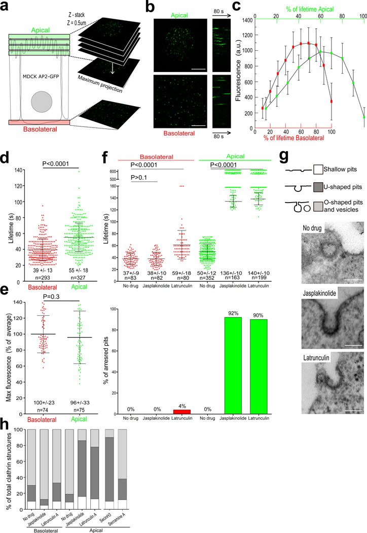
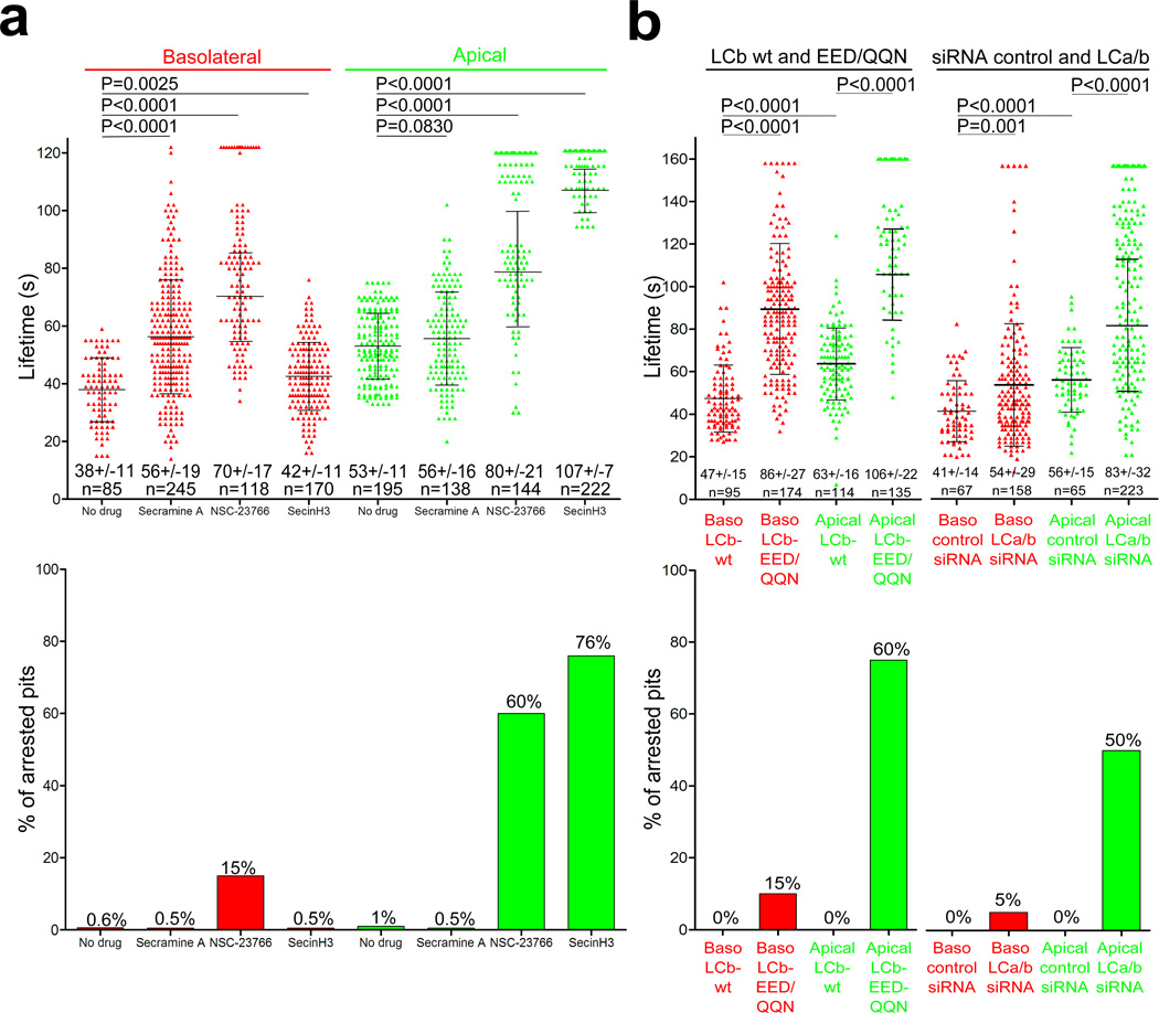
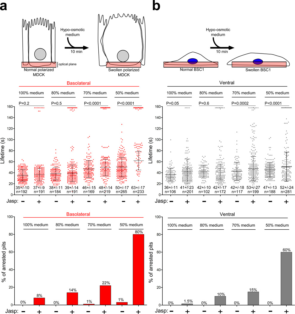
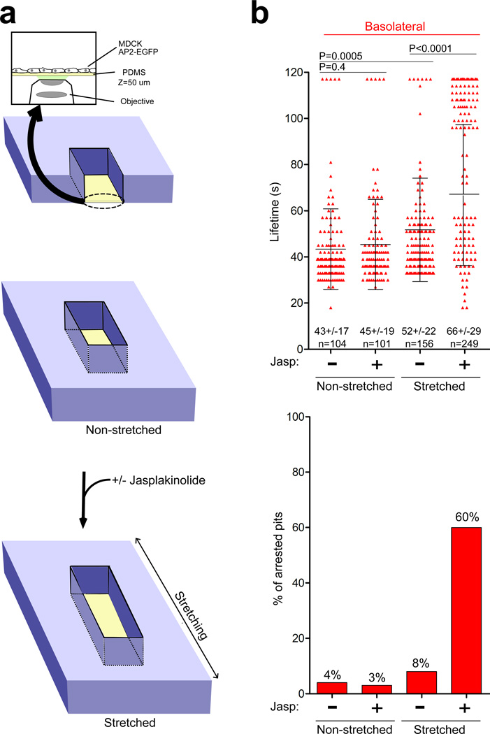
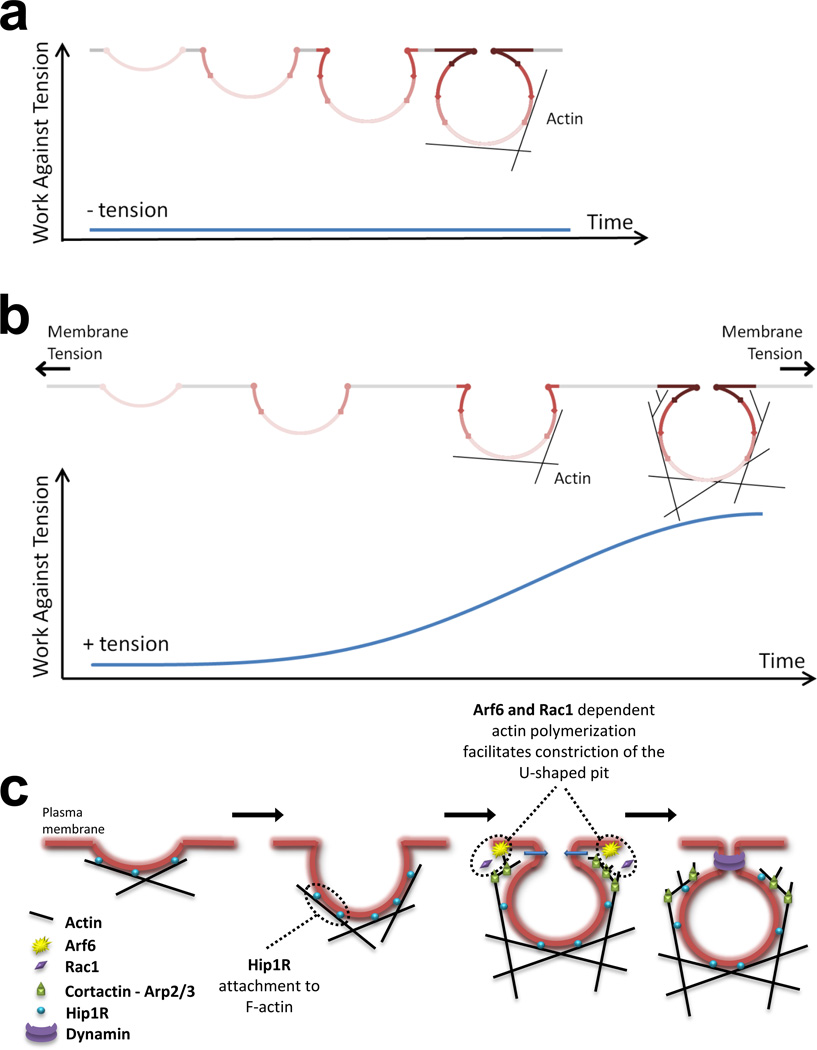
Clathrin-mediated endocytosis is independent of actin dynamics in many circumstances but requires actin polymerization in others. We show that membrane tension determines the actin dependence of clathrin-coat assembly. As found previously, clathrin assembly supports formation of mature coated pits in the absence of actin polymerization on both dorsal and ventral surfaces of non-polarized mammalian cells, and also on basolateral surfaces of polarized cells. Actin engagement is necessary, however, to complete membrane deformation into a coated pit on apical surfaces of polarized cells and, more generally, on the surface of any cell in which the plasma membrane is under tension from osmotic swelling or mechanical stretching. We use these observations to alter actin dependence experimentally and show that resistance of the membrane to propagation of the clathrin lattice determines the distinction between 'actin dependent and 'actin independent'. We also find that light-chain-bound Hip1R mediates actin engagement. These data thus provide a unifying explanation for the role of actin dynamics in coated-pit budding.


Figures

Figure 1
Figure 1. Formation of endocytic coated pits and vesicles at the apical and basolateral surfaces of polarized MDCK cells
(a) Imaging procedures used to visualize the dynamics of pit formation at the apical and basolateral surfaces of polarized MDCK cells. Movies from the dome-like apical surface are from 3D time series acquired at 2 s intervals from 3–5 serial optical sections spaced by 0.5 µm and using 100 ms exposures; 2-D time series were created from maximum intensity z-projection sets. Movies from the basolateral surface are 2-D time series acquired at 2 s intervals from a single optical section. (b) Snapshot from a maximum intensity projection and representative kymograph of coated pit formation at the apical and basolateral surfaces from the same polarized MDCK cell; AP-2 labeled with σ2-EGFP; scale bar, 5 µm. (c) Average fluorescence intensity of AP-2 structures forming at the apical and basolateral surfaces of polarized MDCK cells (N=3), normalized to the lifetime of each individual pit analyzed (% of lifetime). Each point represents average +/− s. d. (d) Scatter plot of individual lifetimes of coated structures from 7 polarized MDCK cells. Each data set represents average +/− s. d. N is the number of objects analyzed. Statistical significances for lifetime differences are shown. (e) Scatter plot of individual maximum fluorescence intensities for coated structures. (f) Scatter plots of lifetimes for individual AP-2 spots at the apical and basolateral surfaces of polarized MDCK cells stably expressing σ2-EGFP in the absence or presence of jasplakinolide or latrunculin A. The upper and lower data sets are from distinct time series of 10 and 2.5 min in duration, respectively. Bottom, fraction of AP-2 objects with longer duration than the time series (% arrested pits). (g) Morphological analysis of clathrin coated structures on the apical surface of polarized MDCK cells treated with jasplakinolide, latrunculin A, SecinH3 or Secramine A. Representative electron microscopy images of the most abundant clathrin-coated pit profiles. Scale bar, 100 nm. The coated structures were classified as shallow, U-shaped or nearly-mature Ω- and fully-mature O-shaped vesicles. (h) Relative frequency of profiles in about 45 cells per condition.
Figure 2
Figure 2. Disruption of apical coat formation by pharmacological interference with the small GTPases Rac1 and Arf6 and interference with the function of clathrin light chains
(a) Top, scatter plots of lifetimes for individual AP-2 spots. Polarized MDCK cells stably expressing σ2-EGFP were treated for 10 min prior to imaging with Secramine A, NSC-23766 or SecinH3, small molecule inhibitors of Cdc42, Rac1 and Arf6, respectively. Each data set represents average +/− s. d for objects whose duration was fully included in the time-series. N is the number of objects analyzed. Statistical significances for the differences in lifetimes are shown. Bottom, fraction of AP-2 objects with longer duration than the time series. (b) Top, scatter plots of lifetimes for individual AP-2 spots imaged at the apical and basolateral surfaces of polarized MDCK cells stably expressing σ2-EGFP and transiently expressing wild type (LCb-wt) or mutant (LCb-EED/QQN) clathrin light chain B fused to cherry (left part) or depleted of both clathrin light chains by siRNA (right part). Each data set represents average +/− s. d. N is the number of objects analyzed. Bottom, fraction of AP-2 objects with longer duration than the 160 s time series.
Figure 3
Figure 3. Actin dependence for endocytic coat formation in cells swelled by hypo-osmotic treatment. Polarized MDCK or non-polarized BSC1 cells incubated for 10 min in serially diluted medium (from 100% to 50%) of decreasing osmolarity, ranging from 311 to 174 mOsm were analyzed for the effects of altering actin dynamics on the lifetimes of their AP-2 coated structures. Each data set represents average +/− s. d.; N, number of objects analyzed
(a) Top, schematic representation of polarized MDCK cells stably expressing σ2-EGFP exposed to hypo-osmotic medium. Middle, scatter plots of lifetimes for individual basolateral AP-2 spots of cells exposed for 10 min to hypoosmotic media in the absence and presence of jasplakinolide. Bottom, fraction of AP-2 objects with longer duration than the time series. (b) Top, schematic representation of BSC1 cells stably expressing σ2-EGFP exposed to hypoosmotic medium. Middle, scatter plots of lifetimes for individual ventral AP-2 spots of cells exposed for 10 min to hypoosmotic media in the absence and presence of jasplakinolide. Bottom, fraction of AP-2 objects with longer duration than the time series.
Figure 4
Figure 4. Actin dependence of endocytic coat formation in mechanically stretched cells
(a) Schematic representation of the device used to image mechanically stretched, non-polarized MDCK cells. An optically clear, stretchable silicon holder made of polydimethylsiloxane (PDMS), about 50 µm thick, which could make contact with the oil above a 63x objective lens was placed at the bottom of the stretching device. Cells were grown for 24 h on the PDMS surface pre-coated with fibronectin and then imaged by spinning disk confocal microscopy; the spherical aberration correction device was essential for detecting the diffraction-limited coated structures containing AP2-EGFP. (b) Dynamics of coated pits before and after ~ 25% linear stretching. Scatter plots for lifetimes of individual AP-2 spots from the ventral surface of cells subjected to controlled stretching in the presence or absence of jasplakinolide. Each data set represents average +/− s. d. N is the number of objects analyzed. Bottom, fraction of AP-2 objects with longer duration than the time series.
Figure 5
Figure 5. Model depicting the role of actin polymerization during the formation of endocytic clathrin coated pits
(a) Under non-stringent conditions and low membrane tension, assembly of the clathrin coat is sufficient to deform the membrane into a tightly constricted coated pit. (b) Under more stringent conditions of high membrane tension, clathrin assembly is not sufficient and membrane invagination stalls; actin polymerization then provides the additional work needed to complete membrane bending. Hip1R links the assembling clathrin coat to actin polymers, and if sufficient time is allowed, then assembly of short-branched actin rescues the stalled coat. The two main forces resisting membrane deformation are bending and tension. The bending work per unit area depends inversely on the curvature and hence is uniform for a spherical or nearly spherical vesicle. The work done against a constant membrane tension depends on the net increase in membrane area -- i.e., the area of the invaginated membrane minus the area of the opening it covers. The plot represents the cumulative work required to counteract membrane tension, and does not include the work required to create the membrane vesicle. The cumulative work was calculated according to W = π r2 T (1 - cosα), where T = membrane tension and α = angle (radians) between the pole of the budding pit and the position at which the curved pit intersects the plane of the plasma membrane (see Meethods). When the neck begins to constrict (α = π/2), the area of the opening decreases and the net increase in area rises sharply. When the pit is complete, α =π. (c) Hip1R links actin filaments with the clathrin coat by interactions with F-actin and clathrin light chains. Branched actin filaments grow towards the plasma membrane via new filament assembly at the barb-end of the stabilized actin filaments; this grow relies on Arp2/3 stimulation, mediated by cortactin and by small GTPases such as Arf6 and Rac1.

References

    1. Ehrlich M, et al. Endocytosis by random initiation and stabilization of clathrin-coated pits. Cell. 2004;118:591–605. - PubMed
    1. Conibear E. Converging views of endocytosis in yeast and mammals. Curr Opin Cell Biol. 2010;22:513–518. - PubMed
    1. Kaksonen M, Toret CP, Drubin DG. A modular design for the clathrin- and actin-mediated endocytosis machinery. Cell. 2005;123:305–320. - PubMed
    1. Merrifield CJ, Feldman ME, Wan L, Almers W. Imaging actin and dynamin recruitment during invagination of single clathrin-coated pits. Nature cell biology. 2002;4:691–698. - PubMed
    1. Boucrot E, Saffarian S, Zhang R, Kirchhausen T. Roles of AP-2 in clathrin-mediated endocytosis. PloS one. 2010;5:e10597. - PMC - PubMed

Publication types

MeSH terms