. 2011 Aug 14;13(10):1244-51.
doi: 10.1038/ncb2313.

Notch post-translationally regulates β-catenin protein in stem and progenitor cells

Affiliations

Notch post-translationally regulates β-catenin protein in stem and progenitor cells

Chulan Kwon et al. Nat Cell Biol. .

Abstract

Cellular decisions of self-renewal or differentiation arise from integration and reciprocal titration of numerous regulatory networks. Notch and Wnt/β-catenin signalling often intersect in stem and progenitor cells and regulate each other transcriptionally. The biological outcome of signalling through each pathway often depends on the context and timing as cells progress through stages of differentiation. Here, we show that membrane-bound Notch physically associates with unphosphorylated (active) β-catenin in stem and colon cancer cells and negatively regulates post-translational accumulation of active β-catenin protein. Notch-dependent regulation of β-catenin protein did not require ligand-dependent membrane cleavage of Notch or the glycogen synthase kinase-3β-dependent activity of the β-catenin destruction complex. It did, however, require the endocytic adaptor protein Numb and lysosomal activity. This study reveals a previously unrecognized function of Notch in negatively titrating active β-catenin protein levels in stem and progenitor cells.


Conflict of interest statement

Competing interests: D.S. serves on the scientific advisory board of iPierian Inc and RegeneRx Pharmaceuticals.

Figures

Figure 1
Figure 1
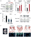
Notch Negatively Regulates Active β-Catenin in Stem Cells Independently of RBP-J. a, Western analysis of ESCs transfected with control or Notch1 (N1) siRNA with active (Act), Phospho (Ser37), or total β-Catenin antibodies that detect N-terminal-dephosphorylated β-Catenin. b, c, Relative β-Catenin/TCF-directed luciferase activity in ESCs (b) or neural stem cells (NSCs) (c) transfected with control siRNA or siRNA against Notch1 or Notch1–4. β-Catenin/TCF activity was measured by co-transfecting cells with a luciferase reporter downstream of multiple TCF binding sites (Topflash). A mutant reporter (Fopflash) exhibited negligible activity in all luciferase assays done in this study. d, Relative RBP-J expression levels by qPCR in ESCs after transfection with control or RBP-J siRNA, determined by qPCR. e, Western analysis of ESCs transfected with control or RBP-J siRNA (50 or 100 nM) with Act β-Cat antibodies. f, Transverse sections of control, Notch1 knockout (KO) (IslCre; Notch1tm2Rko(ex3)loxP) or RBP-J KO (IslCr; RBP-Jflox/flox) embryos stained with H&E (top) or Isl1 antibody (green, bottom) at embryonic day 9.5, at level of outflow tract (ot). Asterisks indicate precardiac mesoderm containing cardiac progenitor cells. Dapi (blue) was used to counterstain the nuclei. The cutting plane is indicated by a dotted line (left). Scale bars, 100 μm. All luciferase values were normalized to Renilla activity (mean±s. d.; n = 4; *P < 0.01). P values were determined using two-tailed Student’s t-test, type II (see Methods). Gapdh antibody was used as a loading control. Numbers on Western blots correspond to relative quantification. h, head; ht, heart tube; Con, control; N1KD, Notch1 siRNA; N1–4KD, Notch 1–4 siRNA.
Figure 2
Figure 2
Notch1 Negatively Regulates Active β-Catenin in ESCs and Physically Interacts with β-Catenin. a, Relative expression of β-Catenin and Cyclin D1 mRNA in ESCs transfected with control or Notch1 siRNA (100 nM), determined by qPCR. b, Western analysis of ESCs transfected with control or Notch1 intracellular domain (N1ICD) (100 or 300 ng) and cultured with BIO. GAPDH antibody was used as a loading control. c, Relative β-Catenin/TCF luciferase activity of BIO-treated ESCs transfected with control or N1ICD +/− MAML or RBP-J siRNA. d, Relative β-Catenin/TCF luciferase activity of ESCs transfected with control or Notch1 siRNA and cultured with or without BIO e, f, Transverse sections of control, IslCre;β-catenin(ex3)loxP (Act-β-Cat) or IslCre;Gt(ROSA)26Sortm1(Notch1)Dam/J (Act-β-Cat; N1ICD overexpression) embryos at embryonic day 9.5, stained with H&E (e) or β-Catenin antibody (red, f). Asterisks indicate precardiac mesoderm containing cardiac progenitor cells (e). Scale bars, 100 μm (e) or 25 μm (f). DAPI (blue) was used to counterstain the nuclei (f). nt, neural tube; ot, outflow tract; pe, pharyngeal endoderm; ec pharyngeal ectoderm; pm, precardiac mesoderm. g, h, ESCs treated with or without BIO (g) or SW480 (human colon cancer) cells (g, h) were transfected with expression constructs for Myc (−) or N1ICD-Myc (+), immunoprecipitated (IP) with anti-Myc antibody and immunoblotted (IB) with β-Catenin antibody recognizing its C-terminus (g), dephosphorylated (active) form, or the phosphorylated N-terminus (h). Notch expression was detected with anti-Myc antibody (g). i, Schematic representation of Notch1 deletion constructs and their interactions with β-Catenin. j, Co-IP of BIO-treated ESCs with Notch1 deletion constructs shown in (i) using antibodies indicated. Arrowheads indicate Notch1 expression. k, Relative β-Catenin/TCF activity of BIO-treated ESCs transfected with Notch constructs shown in (i). TM (transmembrane domain), R (RAM domain), ANK (Ankyrin repeats), TA (transactivation domain), P (PEST domain). BIO was used at 2 μM. All qPCR or luciferase values were normalized to Gapdh or Renilla activity, respectively. (mean± s. d.; n = 4; *P < 0.01). P values were determined using two-tailed Student’s t-test, type II (see Methods). Numbers on Western blots correspond to relative quantification. Con, control; N1KD, Notch1 siRNA.
Figure 3
Figure 3
Membrane-Bound Notch1 Negatively Regulates Active β-Catenin Levels through Numb and Numb-like in Stem Cells. a, Schematic representation of a cleavage site–mutated tethered form of Notch1 (V1774L, top) and Western analysis of ESCs transfected with Myc-tagged wild type Notch1 or tethered Notch1 (V1774L) and blotted with anti-myc antibody (bottom), showing lack of cleaved protein band. b, Relative RBP-J responsive luciferase activity of mESCs transfected with control, tethered Notch1 or N1ICD. c, Relative β-Catenin/TCF luciferase activity of ESCs transfected with control or tethered Notch1 (V1774L) construct shown in (a) or treated with Dkk1 (50 ng/ml). d, BIO-treated ESCs transfected with control or tethered Notch1-myc constructs immunoprecipitated (IP) with anti-Myc antibody and immunoblotted (IB) with β-Catenin antibody. Notch1 expression was detected with anti-Myc antibody. e, Western analysis of active or total β-Catenin in BIO-treated ESCs transfected with control or tethered Notch1. f, % of Brachyury-GFP+ cells after 3 days of differentiation of mouse ESCs with tethered Notch (V1774L) or control in the presence or absence of BIO (0.5 μM) (mean± s. d.; n = 4; *P < 0.01). g, Relative β-Catenin/TCF luciferase activity of ESCs or NSCs treated with increasing doses of DAPT, a γ-secretase inhibitor, for 72–96 h. h, Western analysis of active β-Catenin in mouse or human ESCs, NSCs, or bone marrow mesenchymal stem cells (MSCs) treated with increasing doses (0, 25, 50 or 100 μM) of DAPT for 72–96 h. i, % of Brachyury-GFP+ cells after 3.5 days of differentiation of mouse ESCs with control or DAPT (mean± s. d.; n = 4; *P < 0.01).. j, Relative β-Catenin/TCF luciferase activity of ESCs treated with control or Batimastat, an α-secretase inhibitor. k, Relative β-Catenin/TCF luciferase activity of wild type ESCs (control) or ESCs with ligand-binding site-deleted Notch1 (Notch1lbd/lbd) treated with Wnt3a. All luciferase values were normalized to Renilla activity (mean± s. d.; n = 4; *P < 0.01). P values were determined using two-tailed Student’s t-test, type II (see Methods). Gapdh antibody was used as a loading control. Numbers on Western blots correspond to relative quantification. BIO was used at 2 μM. Con, control.
Figure 4
Figure 4
Notch-Mediated Degradation of β-Catenin Requires Numb and Lysosomal Activity. a, Human colon cancer cells (SW480) transfected with pcDNA-Myc or tethered Notch (V1774L)-Myc constructs, immunoprecipitated (IP) with anti-Myc antibody and immunoblotted (IB) with anti-Numb antibody. Expression of tethered Notch was detected with anti-Myc antibody; expression of pcDNA-myc was confirmed by PCR. b, Relative β-Catenin/TCF luciferase activity of ESCs transfected with control, N1ICD or tethered Notch (V1774L) in the presence or absence of Numb/Numbl siRNA and cultured in BIO for 72 h. c, Western analysis of active β-Catenin in ESCs transfected with control or tethered Notch (V1774L) in the presence or absence of Numb/Numbl siRNA. d, Western analysis of active β-Catenin in ESCs treated with control or DAPT in the presence or absence of Bafilomycin A1, which inhibits lysosomal activity. All luciferase values were normalized to Renilla activity (mean± s. d.; n = 4; *P < 0.01; NS, not significant). P values were determined using two-tailed Student’s t-test, type II (see Methods). Gapdh antibody was used as a loading control. Numbers on Western blots correspond to relative quantification. Con, control.
Figure 5
Figure 5
γ-Secretase Inhibitors (GSIs) Suppress Expansion of Human Colon Cancer Cells by Blocking Notch Cleavage. a, Western analysis of active β-Catenin in SW480 human colon cancer cells transfected with control or siRNA against Notch1–4 (N1-4KD, 100 nM each). b, Relative β-Catenin/TCF luciferase activity of SW480 cells treated with increasing doses of DAPT for 96 h. c, Western analysis of β-Catenin levels in SW480 and a second colon cancer cell line, HT-29, treated with increasing doses (0, 25, 50 or 100 μM) of DAPT for 96 h. d, Relative number of SW480 cells treated with DAPT (50 or 100 μM) for 72 h (mean± s. d.; n = 4; *P < 0.01). e, Western analysis of active β-Catenin levels in SW480 cells with increasing DAPT in the presence or absence of proteasome inhibitor (PI) MG-132 (5 nM) for 72 h. Fewer PI-treated cells were loaded in the right panel since they exhibit higher levels of β-Catenin. f, Notch/RBP-J luciferase reporter activity (multimerized RBP-J binding sites) of SW480 cells treated with increasing doses of ibuprofen. g, Relative β-Catenin/TCF luciferase activity of SW480 cells treated with Ibuprofen for 72 h. h, Western analysis of active β-Catenin in SW480 cells treated with Ibuprofen for 72 h. i, Western analysis of active β-Catenin in SW480 cells treated with control or Ibuprofen and transfected with Notch1–4 (100 nM each) siRNA. GAPDH antibody was used as a loading control. All luciferase values were normalized to Renilla activity (mean± s. d.; n = 4; *P < 0.01). P values were determined using two-tailed Student’s t-test, type II (see Methods). Numbers on Western blots correspond to relative quantification. Con, control; N1–4KD, Notch 1–4 siRNA. j, Model for Post-Translational Regulation of β-Catenin Protein by Notch. In the absence of Wnt, the destruction complex of Axin, APC and GSK3β phosphorylates β-Catenin, leading to its proteasomal degradation (left). When the destruction complex is inactivated by Wnts, dephosphorylated (active) β-Catenin functions as a transcriptional activator with TCF/LEF. We show that active β-Catenin protein levels can be negatively regulated by interaction with Notch in a Numb-dependent manner, involving the lysosome. Notch-mediated degradation of β-Catenin is independent of the APC-dependent destruction complex.

References

    1. Logan CY, Nusse R. The Wnt signaling pathway in development and disease. Annu Rev Cell Dev Biol. 2004;20:781–810. - PubMed
    1. Radtke F, Clevers H. Self-renewal and cancer of the gut: two sides of a coin. Science. 2005;307:1904–1909. - PubMed
    1. Hayward P, Kalmar T, Arias AM. Wnt/Notch signalling and information processing during development. Development. 2008;135:411–424. - PubMed
    1. Artavanis-Tsakonas S, Rand MD, Lake RJ. Notch signaling: cell fate control and signal integration in development. Science. 1999;284:770–776. - PubMed
    1. Kwon C, et al. A regulatory pathway involving Notch1/beta-catenin/Isl1 determines cardiac progenitor cell fate. Nat Cell Biol. 2009;11:951–957. - PMC - PubMed

Publication types

MeSH terms