

α-Synuclein occurs physiologically as a helically folded tetramer that resists aggregation
- PMID: 21841800
- PMCID: PMC3166366
- DOI: 10.1038/nature10324
α-Synuclein occurs physiologically as a helically folded tetramer that resists aggregation
Abstract
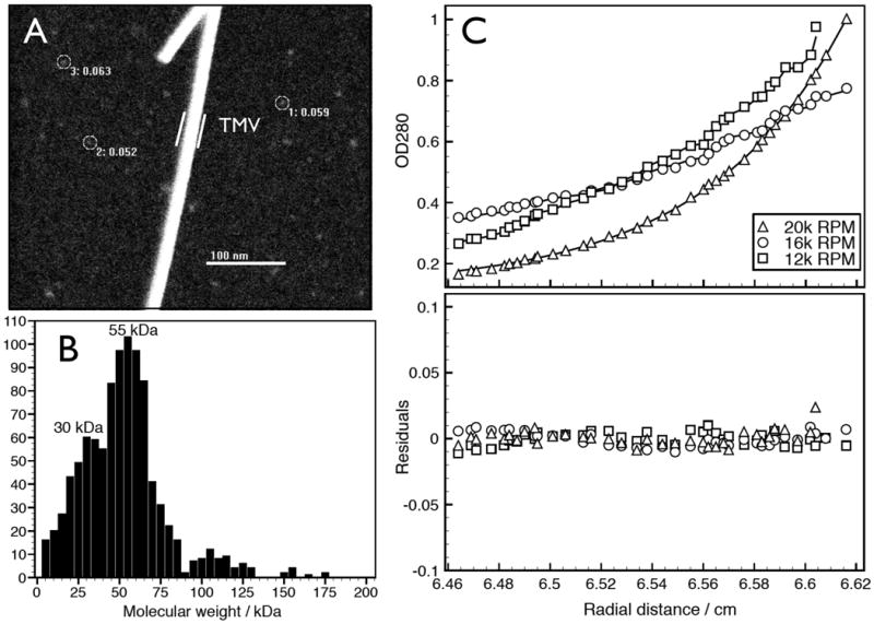
Parkinson's disease is the second most common neurodegenerative disorder. Growing evidence indicates a causative role of misfolded forms of the protein α-synuclein in the pathogenesis of Parkinson's disease. Intraneuronal aggregates of α-synuclein occur in Lewy bodies and Lewy neurites, the cytopathological hallmarks of Parkinson's disease and related disorders called synucleinopathies. α-Synuclein has long been defined as a 'natively unfolded' monomer of about 14 kDa (ref. 6) that is believed to acquire α-helical secondary structure only upon binding to lipid vesicles. This concept derives from the widespread use of recombinant bacterial expression protocols for in vitro studies, and of overexpression, sample heating and/or denaturing gels for cell culture and tissue studies. In contrast, we report that endogenous α-synuclein isolated and analysed under non-denaturing conditions from neuronal and non-neuronal cell lines, brain tissue and living human cells occurs in large part as a folded tetramer of about 58 kDa. Several methods, including analytical ultracentrifugation, scanning transmission electron microscopy and in vitro cell crosslinking confirmed the occurrence of the tetramer. Native, cell-derived α-synuclein showed α-helical structure without lipid addition and had much greater lipid-binding capacity than the recombinant α-synuclein studied heretofore. Whereas recombinantly expressed monomers readily aggregated into amyloid-like fibrils in vitro, native human tetramers underwent little or no amyloid-like aggregation. On the basis of these findings, we propose that destabilization of the helically folded tetramer precedes α-synuclein misfolding and aggregation in Parkinson's disease and other human synucleinopathies, and that small molecules that stabilize the physiological tetramer could reduce α-synuclein pathogenicity.
Figures


Comment in
-
Neurodegenerative disease: α-synuclein gets a new look.Nat Rev Neurosci. 2011 Sep 2;12(10):550. doi: 10.1038/nrn3106. Nat Rev Neurosci. 2011. PMID: 21886184 No abstract available.
-
The true nature of α-synuclein unmasked!Mov Disord. 2011 Nov;26(13):2324. doi: 10.1002/mds.24009. Mov Disord. 2011. PMID: 22319787 No abstract available.
-
Properties of native brain α-synuclein.Nature. 2013 Jun 13;498(7453):E4-6; discussion E6-7. doi: 10.1038/nature12125. Nature. 2013. PMID: 23765500 Free PMC article. No abstract available.
References
-
- Obeso JA, et al. Missing pieces in the Parkinson's disease puzzle. Nat Med. 2010;16:653–661. - PubMed
-
- Tong J, et al. Brain {alpha}-synuclein accumulation in multiple system atrophy, Parkinson's disease and progressive supranuclear palsy: a comparative investigation. Brain. 2010;133:172–188. - PubMed
-
- Spillantini MG, et al. Alpha-synuclein in Lewy bodies. Nature. 1997;388:839–840. - PubMed
Publication types
MeSH terms
Substances
Grants and funding
LinkOut - more resources
Full Text Sources
Other Literature Sources
Molecular Biology Databases
