

Effects of di-n-butyl phthalate on male rat reproduction following pubertal exposure
- PMID: 21841806
- PMCID: PMC3739579
- DOI: 10.1038/aja.2011.76
Effects of di-n-butyl phthalate on male rat reproduction following pubertal exposure
Abstract
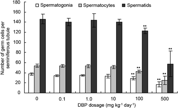
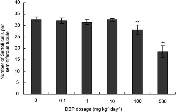
Di-n-butyl phthalate (DBP) is an endocrine-disrupting chemical that has the potential to affect male reproduction. However, the reproductive effects of low-dose DBP are still not well known, especially at the molecular level. In the present study, pubertal male Sprague-Dawley rats were orally administered DBP at a wide range of doses (0.1, 1.0, 10, 100 and 500 mg kg⁻¹ day⁻¹) for 30 days. The selected end points included reproductive organ weights, testicular histopathology and serum hormonal levels. Additionally, proteomic analysis was performed to identify proteins that are differentially expressed as a result of exposure to DBP at low doses (0.1, 1.0 and 10 mg kg⁻¹ day⁻¹). Toxic effects were observed in the high-dose groups, including anomalous development of testes and epididymides, severe atrophy of seminiferous tubules, loss of spermatogenesis and abnormal levels of serum hormones. Treatment with low doses of DBP seemed to exert a 'stimulative effect' on the serum hormones. Proteomics analysis of rat testes showed 20 differentially expressed proteins. Among these proteins, alterations in the expression of HnRNPA2/B1, vimentin and superoxide dismutase 1 (SOD1) were further confirmed by Western blot and immunohistochemistry. Taken together, we conclude that high doses of DBP led to testicular toxicity, and low doses of DBP led to changes in the expression of proteins involved in spermatogenesis as well as changes in the number and function of Sertoli and Leydig cells, although no obvious morphological changes appeared. The identification of these differentially expressed proteins provides important information about the mechanisms underlying the effects of DBP on male rat reproduction.
Figures


Similar articles
-
Di-n-butyl phthalate prompts interruption of spermatogenesis, steroidogenesis, and fertility associated with increased testicular oxidative stress in adult male rats.Environ Sci Pollut Res Int. 2017 Aug;24(22):18563-18574. doi: 10.1007/s11356-017-9478-3. Epub 2017 Jun 24. Environ Sci Pollut Res Int. 2017. PMID: 28646317
-
NTP technical report on the toxicity studies of Dibutyl Phthalate (CAS No. 84-74-2) Administered in Feed to F344/N Rats and B6C3F1 Mice.Toxic Rep Ser. 1995 Apr;30:1-G5. Toxic Rep Ser. 1995. PMID: 12209194
-
The effects on steroidogenesis and histopathology of adult male Japanese quails (Coturnix coturnix japonica) testis following pre-pubertal exposure to di(n-butyl) phthalate (DBP).Comp Biochem Physiol C Toxicol Pharmacol. 2014 Nov;166:24-33. doi: 10.1016/j.cbpc.2014.06.005. Epub 2014 Jun 28. Comp Biochem Physiol C Toxicol Pharmacol. 2014. PMID: 24983780
-
The overview of current evidence on the reproductive toxicity of dibutyl phthalate.Int J Occup Med Environ Health. 2021 Jan 7;34(1):15-37. doi: 10.13075/ijomeh.1896.01658. Epub 2020 Nov 13. Int J Occup Med Environ Health. 2021. PMID: 33223541 Review.
-
Effects of di-n-butyl phthalate (DBP) on male reproductive development in the rat: implications for human risk assessment.Food Chem Toxicol. 2000;38(1 Suppl):S97-9. doi: 10.1016/s0278-6915(99)00128-3. Food Chem Toxicol. 2000. PMID: 10717378 Review.
Cited by
-
Di-n-butyl phthalate disrupts the expression of genes involved in cell cycle and apoptotic pathways in mouse ovarian antral follicles.Biol Reprod. 2013 Jan 31;88(1):23. doi: 10.1095/biolreprod.112.105122. Print 2013 Jan. Biol Reprod. 2013. PMID: 23242528 Free PMC article.
-
Di-n-butyl phthalate prompts interruption of spermatogenesis, steroidogenesis, and fertility associated with increased testicular oxidative stress in adult male rats.Environ Sci Pollut Res Int. 2017 Aug;24(22):18563-18574. doi: 10.1007/s11356-017-9478-3. Epub 2017 Jun 24. Environ Sci Pollut Res Int. 2017. PMID: 28646317
-
Use of the Adverse Outcome Pathway (AOP) framework to evaluate species concordance and human relevance of Dibutyl phthalate (DBP)-induced male reproductive toxicity.Reprod Toxicol. 2020 Sep;96:445-458. doi: 10.1016/j.reprotox.2019.06.009. Epub 2019 Jun 28. Reprod Toxicol. 2020. PMID: 31260805 Free PMC article. Review.
-
Di-n-butyl phthalate epigenetically induces reproductive toxicity via the PTEN/AKT pathway.Cell Death Dis. 2019 Apr 5;10(4):307. doi: 10.1038/s41419-019-1547-8. Cell Death Dis. 2019. PMID: 30952838 Free PMC article.
-
Smad2/3 Upregulates the Expression of Vimentin and Affects Its Distribution in DBP-Exposed Sertoli Cells.PPAR Res. 2015;2015:489314. doi: 10.1155/2015/489314. Epub 2015 Dec 24. PPAR Res. 2015. PMID: 26819576 Free PMC article.
References
-
- Bosnir J, Puntarić D, Skes I, Klarić M, Simić S, et al. Migration of phthalates from plastic products to model solutions. Coll Antropol. 2003;27 Suppl 1:23–30. - PubMed
-
- Kavlock R, Boekelheide K, Chapin R, Cunningham M. NTP center for the evaluation of risks to human reproduction: phthalates expert panel report on the reproductive and developmental toxicity of di(2-ethylhexyl) phthalate. Reprod Toxicol. 2002;16:529–653. - PubMed
-
- Gray LE Jr, Wolf C, Lambright C, Mann P, Price M, et al. Administration of potentially antiandrogenic pesticides (procymidone, linuron, iprodione, chlozolinate, p,p'-DDE, and ketoconazole) and toxic substances (dibutyl- and diethylhexyl phthalate, PCB 169, and ethane dimethane sulphonate) during sexual differentiation produces diverse profiles of reproductive malformations in the male rat. Toxicol Ind Health. 1999;15:94–118. - PubMed
-
- Mylchreest E, Sar M, Cattley RC, Foster PM. Disruption of androgen-regulated male reproductive development by di(n-butyl) phthalate during late gestation in rats is different from flutamide. Toxicol Appl Pharmacol. 1999;156:81–95. - PubMed
Publication types
MeSH terms
Substances
LinkOut - more resources
Full Text Sources
Miscellaneous
