Coronary slow flow and acute coronary syndrome in a patient with spinal cord injury
- PMID: 21841878
- PMCID: PMC3147186
Coronary slow flow and acute coronary syndrome in a patient with spinal cord injury
Abstract
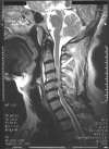
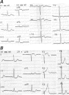
We report the case of a 55-year-old man who presented with acute coronary syndrome due to coronary slow flow after spinal cord injury. Data regarding the causes and clinical manifestations of coronary slow flow are inconclusive, but the autonomic nervous system is believed to be at least a contributing factor. The predominant vagal activity causes vasodilation and hemostasis, which can lead to acute coronary syndrome. We hereby call attention to hyperactive parasympathetic tonicity, which can lead to coronary slow flow and acute coronary syndrome in acute spinal cord injury patients.
Keywords: Acute coronary syndrome; autonomic dysreflexia; coronary circulation; coronary slow flow; spinal cord injuries/complications.
Figures


Similar articles
-
The rare complication of Behcet's syndrome: concomitance of coronary slow flow with acute coronary syndrome.BMJ Case Rep. 2012 Jul 27;2012:bcr2012006388. doi: 10.1136/bcr-2012-006388. BMJ Case Rep. 2012. PMID: 22847567 Free PMC article. No abstract available.
-
Clinical manifestations of slow coronary flow from acute coronary syndrome to serious arrhythmias.Cardiol J. 2009;16(5):462-8. Cardiol J. 2009. PMID: 19753527
-
Perforated balloon technique: A simple and handy technique to combat no-reflow phenomenon in coronary system.Catheter Cardiovasc Interv. 2018 Nov 1;92(5):890-894. doi: 10.1002/ccd.27477. Epub 2017 Dec 27. Catheter Cardiovasc Interv. 2018. PMID: 29280545
-
Slow Coronary Flow: Pathophysiology, Clinical Implications, and Therapeutic Management.Angiology. 2021 Oct;72(9):808-818. doi: 10.1177/00033197211004390. Epub 2021 Mar 29. Angiology. 2021. PMID: 33779300 Review.
-
The pathogenesis and treatment of no-reflow occurring during percutaneous coronary intervention.Cardiovasc Revasc Med. 2008 Jan-Mar;9(1):56-61. doi: 10.1016/j.carrev.2007.08.005. Cardiovasc Revasc Med. 2008. PMID: 18206640 Review.
References
-
- Sezgin AT, Sigirci A, Barutcu I, Topal E, Sezgin N, Ozdemir R, et al. Vascular endothelial function in patients with slow coronary flow. Coron Artery Dis 2003;14(2):155–61. - PubMed
-
- Duncker DJ, Bache RJ. Regulation of coronary blood flow during exercise. Physiol Rev 2008;88(3):1009–86. - PubMed
-
- Phillips WT, Kiratli BJ, Sarkarati M, Weraarchakul G, Myers J, Franklin BA, et al. Effect of spinal cord injury on the heart and cardiovascular fitness. Curr Probl Cardiol 1998;23(11): 641–716. - PubMed
-
- Feigl EO. Coronary physiology. Physiol Rev 1983;63(1):1–205. - PubMed
-
- Jacobs PL, Mahoney ET, Robbins A, Nash M. Hypokinetic circulation in persons with paraplegia. Med Sci Sports Exerc 2002;34(9):1401–7. - PubMed
Publication types
MeSH terms
Substances
LinkOut - more resources
Full Text Sources
Medical
Miscellaneous
