. 2010 Sep 23:8:e0135.
doi: 10.1199/tab.0135.

The Cryptochrome Blue Light Receptors

Affiliations

The Cryptochrome Blue Light Receptors

Xuhong Yu et al. Arabidopsis Book. .

Abstract

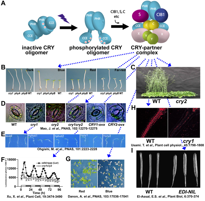
Cryptochromes are photolyase-like blue light receptors originally discovered in Arabidopsis but later found in other plants, microbes, and animals. Arabidopsis has two cryptochromes, CRY1 and CRY2, which mediate primarily blue light inhibition of hypocotyl elongation and photoperiodic control of floral initiation, respectively. In addition, cryptochromes also regulate over a dozen other light responses, including circadian rhythms, tropic growth, stomata opening, guard cell development, root development, bacterial and viral pathogen responses, abiotic stress responses, cell cycles, programmed cell death, apical dominance, fruit and ovule development, seed dormancy, and magnetoreception. Cryptochromes have two domains, the N-terminal PHR (Photolyase-Homologous Region) domain that bind the chromophore FAD (flavin adenine dinucleotide), and the CCE (CRY C-terminal Extension) domain that appears intrinsically unstructured but critical to the function and regulation of cryptochromes. Most cryptochromes accumulate in the nucleus, and they undergo blue light-dependent phosphorylation or ubiquitination. It is hypothesized that photons excite electrons of the flavin molecule, resulting in redox reaction or circular electron shuttle and conformational changes of the photoreceptors. The photoexcited cryptochrome are phosphorylated to adopt an open conformation, which interacts with signaling partner proteins to alter gene expression at both transcriptional and posttranslational levels and consequently the metabolic and developmental programs of plants.


Figures

Figure 1.
Figure 1.
Arabidopsis cryptochromes undergo light-dependent conformational change to interact with multiple signaling proteins and regulate multiple light responses. (A) A model depicting constitutive homodimerization of cryptochrome via the PHR domain, light-dependent phosphorylation (negative charges shown) and changes of conformation (depicted as the disengagement of the PHR and CCE domains), and interaction with signaling proteins, including CIB1, COP1 (C), SPA's (S), and other unidentified proteins (X and Y). This figure was modified from Figure 4D in Yu et al. (2007a) with permission from the National Academy of Sciences, USA. (B–I) Different functions of Arabidopsis cryptochromes are shown by comparisons of different genotypes grown under different light conditions. The respective genotypes or light conditions are indicated underneath. (B) Arabidopsis seedlings grown in the dark, continuous blue, red, or far-red light, showing blue light-specific long hypocotyl phenotype of the cry1 mutant. (C) Arabidopsis wild-type (WT) and the cry2 mutant (cry2) grown in long-day photoperiod, showing late-flowering phenotype of the cry2 mutant. (D–I) Images taken from the published articles (references are indicated underneath) with permission show cryptochromes mediate blue light stimulation of stomata opening (D), random bending of hypocotyls (E), the circadian oscillation of cytoplasmic calcium concentration (F), programmed cell death (of the flu mutant) (G), plastid development (H), and fruit (silique) elongation (I).
Figure 2.
Figure 2.
CRY2 is found in nucleoplasm during interphase but it is associated with chromosomes during mitosis. (A) Fluorescence images of CRY2-GFP (left and center) or GFP-CRY2 (right) fusion proteins taken from mesophyll cells (left) or from shoot apex cells (center and right) of the respective transgenic plants grown in white light. Arrows indicate mitotic cells and CRY2-associated chromosomes. (B) An enlarged image of the boxed area of panel A (center) showing association of CRY2-GFP with condensed chromosomes. (C) The cover of “The Arabidopsis Book”, showing the fusion protein of GFP and the C-terminal region of CRY2 (CCE2) in nuclearplasm (the surrounding cells) or chromosomes (the central cell).
Figure 3.
Figure 3.
Structure and electrostatic surfaces of photolyase and cryptochromes (A) Secondary structure of Synechococcus elongatus photolyase (PDB ID 1QNF) and the PHR domain of Arabidopsis CRY1 (PDB ID 1U3C). (B) Electrostatic surface potentials of SePhotolyase, Arabidopsis CRY-DASH/ CRY3 (PDB ID 2IJG), Arabidopsis CRY1-PHR domain, and Arabidopsis CRY2 (predicted structure by homology modeling). Images were generated using Pymol (http://www.pymol.org).
Figure 4.
Figure 4.
Structure features of Arabidopsis CRY1 (A) Sequence alignment of a conserved region of the FAD-binding pocket of human (HsCRY1 and HsCRY2), Drosophila (DmCRY), and Arabidopsis (AtCRY1 and AtCRY2) cryptochromes. Asterisk indicates D387 of CRY2 and equivalent residues in other cryptochromes. (B) Absorption spectrum of Arabidopsis CRY2 and CRY2D387A mutant proteins, showing the lack of blue light absorption of the CRY2D3S7A mutant protein (panels A and B are from Liu et al., 2008a). (C) The secondary structure of the PHR domain of Arabidopsis CRY1 (PDB ID 1U3C), showing the relative position of FAD (yellow) and the residue D390 (red) of CRY1 that is equivalent to D387 of CRY2. (D) Atomic structure of the region surrounding FAD and D390 of CRY1. (E) Distances (Å) between indicated atoms of D390 and FAD. (F) The local disorder tendency of the CCE domain of CRY2 based on an estimated-amino-acid-pairwise-energy-content analysis by IUPred (http://iupred.enzim.hu/), indicating intrinsically unstructured nature of the CCE domain of CRY2.
Figure 5.
Figure 5.
Oxidoreduction of flavins Five possible redox forms of flavins are shown. R indicates different side groups in different flavins. The two different forms of semiquinone radicals: anion red radical (e.g. FAD·—) and neutral blue radical (e.g. FADH·), and two forms of reduced flavins: protonated hydroquinone (e.g. FADH2) and anionic hydroquinone (e.g. FADH) are shown.
Figure 6.
Figure 6.
Working models of the signal transduction mechanism of Arabidopsis cryptochromes Three possible signaling pathways by which CRY1 (A) or CRY2 (B) may regulate gene expression changes and developments of plants in response to blue light are depicted. (A) CRY1 mediates blue light suppression of the COP1-dependent degradation of known (HY5, HYH, HFR1) or unknown (X) transcription regulators to affect expression of light-regulated genes (LRG's). CRY1 may also directly or indirectly interact with unknown (X) transcription regulators. In addition, CRY1 mediates light entrainment of the circadian clock via unknown mechanisms to gate light control of LRG's. (B) CRY2 undergoes blue light-dependent interaction with the CIB transcription factors to directly stimulate transcription of the target gene FT. In addition, CRY2 also interact with COP1-interacting proteins to suppress COP1-dependent degradation of the CO protein that is a positive regulator of FT transcription.

References

    1. Ahmad M., Cashmore A.R. HY4 gene of A. thaliana encodes a protein with characteristics of a blue-light photoreceptor. Nature. 1993;3668(1):162–166. - PubMed
    1. Ahmad M., Lin C., Cashmore A.R. Mutations throughout an Arabidopsis blue-light photoreceptor impair blue-light-responsive anthocyanin accumulation and inhibition of hypocotyl elongation. Plant J. 1995;88(1):653–658. - PubMed
    1. Ahmad M., Jarillo J.A., Cashmore A.R. Chimeric proteins between cry1 and cry2 arabidopsis blue light photoreceptors indicate overlapping functions and varying protein stability. Plant Cell. 1998a;108(1):197–208. - PMC - PubMed
    1. Ahmad M., Jarillo J.A., Smirnova O., Cashmore A.R. Cryptochrome blue-light photoreceptors of Arabidopsis implicated in phototropism. Nature. 1998b;3928(1):720–723. - PubMed
    1. Ahmad M., Jarillo J.A., Smirnova O., Cashmore A.R. The CRY1 blue light photoreceptor of Arabidopsis interacts with phytochrome A in vitro. Mol Cell. 1998c;18(1):939–948. - PubMed