

Language organization and reorganization in epilepsy
- PMID: 21842185
- PMCID: PMC3193181
- DOI: 10.1007/s11065-011-9180-z
Language organization and reorganization in epilepsy
Abstract
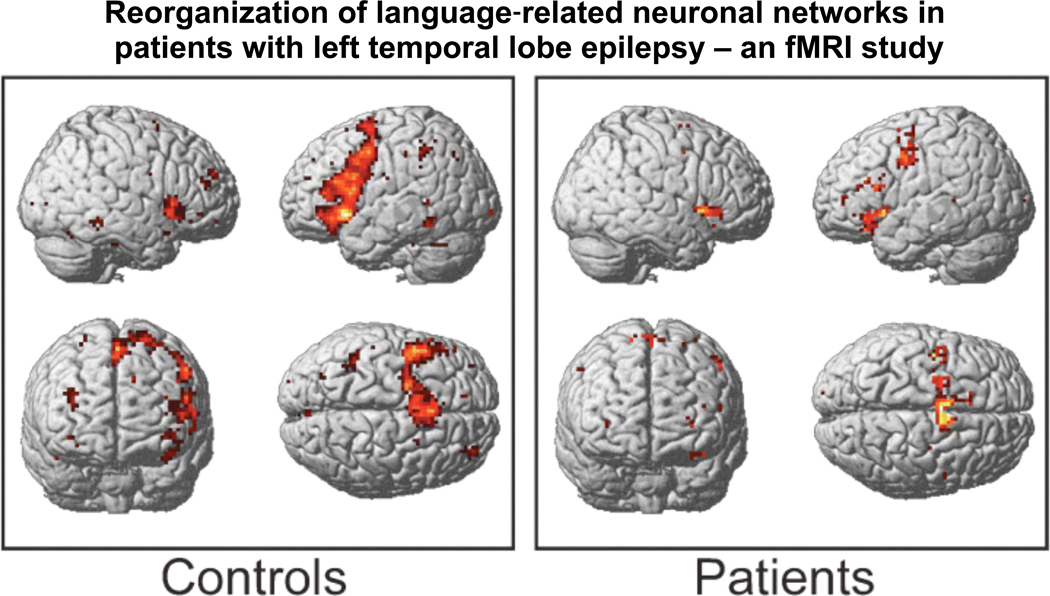
The vast majority of healthy individuals are left hemisphere dominant for language; however, individuals with left hemisphere epilepsy have a higher likelihood of atypical language organization. The cerebral organization of language in epilepsy has been studied with invasive procedures such as Wada testing and electrical cortical stimulation mapping (ESM), and more recently, with noninvasive neuroimaging techniques such as functional magnetic resonance imaging (fMRI). Investigators have used these techniques to explore the influence of unique clinical features inherent in epilepsy that might contribute to the reorganization of language, such as location of seizure onset, age of seizure onset, and extent of interictal epileptiform activity. In this paper, we review the contribution of these and other clinical variables to the lateralization and localization of language in epilepsy, and how these patient-related variables affect the results from these three different, yet complementary methodologies. Unlike the abrupt language changes that occur following acute brain injury with disruption of established language circuits, converging evidence suggests that the chronic nature of epileptic activity can result in a developmental shift of language from the left to the right hemisphere or re-routing of language pathways from traditional to non-traditional areas within the dominant left hemisphere. Clinical variables have been shown to contribute to cerebral language reorganization in the setting of chronic seizure disorders, yet such factors have not been reliable predictors of altered language networks in individual patients, underscoring the need for language lateralization and localization procedures when definitive identification of language cortex is necessary for clinical care.
Conflict of interest statement
Disclosure: The authors report no conflicts of interest
Figures
References
-
- Aarts JH, Binnie CD, Smit AM, Wilkins AJ. Selective cognitive impairment during focal and generalized epileptiform EEG activity. Brain. 1984;107:293–308. - PubMed
-
- Adcock JE, Wise RG, Oxbury JM, Oxbury SM, Matthews PM. Quantitative fMRI assessment of the differences in lateralization of language-related brain activation in patients with temporal lobe epilepsy. Neuroimage. 2003;18:423–438. - PubMed
-
- Balsamo LM, W.D G. The utility of functional mangnetic resonance imaging in epilepsy and language. Current Neurology and Neuroscience Reports. 2002;2:141–149. - PubMed
-
- Belliveau J, Rosen B, Kantor H, Rzedzian RKD, McKinstry R, Vevea J, et al. Functional cerebral imaging by susceptibility-contrast NMR. Magn Reson Med. 1990;14:538–546. - PubMed
-
- Benke T, Koylu B, Visani P, Karner E, Brenneis C, Bartha L, et al. Language lateralization in temporal lobe epilepsy: A comparison between fMRI and the Wada test. Epilepsia. 2006;47(8):1308–1319. - PubMed
Publication types
MeSH terms
Grants and funding
LinkOut - more resources
Full Text Sources
Medical
