In vitro cellular uptake and dimerization of signal transducer and activator of transcription-3 (STAT3) identify the photosensitizing and imaging-potential of isomeric photosensitizers derived from chlorophyll-a and bacteriochlorophyll-a
- PMID: 21842893
- PMCID: PMC3188669
- DOI: 10.1021/jm200805y
In vitro cellular uptake and dimerization of signal transducer and activator of transcription-3 (STAT3) identify the photosensitizing and imaging-potential of isomeric photosensitizers derived from chlorophyll-a and bacteriochlorophyll-a
Abstract
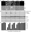
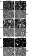
Among the photosensitizers investigated, both ring-D and ring-B reduced chlorins containing the m-iodobenzyloxyethyl group at position-3 and a carboxylic acid functionality at position-17(2) showed the highest uptake by tumor cells and light-dependent photoreaction that correlated with maximal tumor-imaging [positron emission tomography (PET) and fluorescence] and long-term photodynamic therapy (PDT) efficacy in BALB/c mice bearing Colon26 tumors. However, among the ring-D reduced compounds, the isomer containing the 1'-m-iobenzyloxyethyl group at position-3 was more effective than the corresponding 8-(1'-m-iodobenzyloxyethyl) derivative. All photosensitizers showed maximum uptake by tumor tissue 24 h after injection, and the tumors exposed with light at low fluence and fluence rates (128 J/cm(2), 14 mW/cm(2)) produced significantly enhanced tumor eradication than those exposed at higher fluence and fluence rate (135 J/cm(2), 75 mW/cm(2)). Interestingly, dose-dependent cellular uptake of the compounds and light-dependent STAT3 dimerization have emerged as sensitive rapid indicators for PDT efficacy in vitro and in vivo and could be used as in vitro/in vivo biomarkers for evaluating and optimizing the in vivo treatment parameters of the existing and new PDT candidates.
Figures


Similar articles
-
Effect of chirality on cellular uptake, imaging and photodynamic therapy of photosensitizers derived from chlorophyll-a.Bioorg Med Chem. 2015 Jul 1;23(13):3603-17. doi: 10.1016/j.bmc.2015.04.006. Epub 2015 Apr 9. Bioorg Med Chem. 2015. PMID: 25936263 Free PMC article.
-
Multimodality agents for tumor imaging (PET, fluorescence) and photodynamic therapy. A possible "see and treat" approach.J Med Chem. 2005 Oct 6;48(20):6286-95. doi: 10.1021/jm050427m. J Med Chem. 2005. PMID: 16190755
-
Measurement of Cyanine Dye Photobleaching in Photosensitizer Cyanine Dye Conjugates Could Help in Optimizing Light Dosimetry for Improved Photodynamic Therapy of Cancer.Molecules. 2018 Jul 24;23(8):1842. doi: 10.3390/molecules23081842. Molecules. 2018. PMID: 30042350 Free PMC article.
-
Nature: a rich source for developing multifunctional agents. Tumor-imaging and photodynamic therapy.Lasers Surg Med. 2006 Jun;38(5):445-67. doi: 10.1002/lsm.20352. Lasers Surg Med. 2006. PMID: 16788930 Review.
-
Development of bacteriochlorophyll a-based near-infrared photosensitizers conjugated to gold nanoparticles for photodynamic therapy of cancer.Biochemistry (Mosc). 2015 Jun;80(6):752-62. doi: 10.1134/S0006297915060103. Biochemistry (Mosc). 2015. PMID: 26531020 Review.
Cited by
-
Selective Determination of Glutathione Using a Highly Emissive Fluorescent Probe Based on a Pyrrolidine-Fused Chlorin.Molecules. 2023 Jan 5;28(2):568. doi: 10.3390/molecules28020568. Molecules. 2023. PMID: 36677627 Free PMC article.
-
Cell-specific Retention and Action of Pheophorbide-based Photosensitizers in Human Lung Cancer Cells.Photochem Photobiol. 2019 May;95(3):846-859. doi: 10.1111/php.13043. Epub 2018 Nov 28. Photochem Photobiol. 2019. PMID: 30378688 Free PMC article.
-
Pathological Mechanism of Photodynamic Therapy and Photothermal Therapy Based on Nanoparticles.Int J Nanomedicine. 2020 Sep 15;15:6827-6838. doi: 10.2147/IJN.S269321. eCollection 2020. Int J Nanomedicine. 2020. PMID: 32982235 Free PMC article. Review.
-
Monitoring Tumor Hypoxia Using (18)F-FMISO PET and Pharmacokinetics Modeling after Photodynamic Therapy.Sci Rep. 2016 Aug 22;6:31551. doi: 10.1038/srep31551. Sci Rep. 2016. PMID: 27546160 Free PMC article.
-
Theranostics with photodynamic therapy for personalized medicine: to see and to treat.Theranostics. 2023 Oct 9;13(15):5501-5544. doi: 10.7150/thno.87363. eCollection 2023. Theranostics. 2023. PMID: 37908729 Free PMC article. Review.
References
-
- Schrevens L, Lorent N, Dooms C, Vensteenkiste J. The role of PET scan in diagnosis, staging and management of non-small cell lung cancer. The Oncologist. 2004;9:633–643. - PubMed
-
- Evans JW, Peters AM. Gamma camera imaging in malignancy. Eur J Cancer. 2002;38:2157–72. - PubMed
-
- Gambhir SS. Molecular imaging of cancer with PET. Nat Rev Cancer. 2002;2:683–693. - PubMed
-
- Ametamey SM, Honer, Schubiger PA. Molecular Imaging and PET. Chem Rev. 2008;108:1501–1516. - PubMed
-
- Jogoda E, Lang L, Caraco C, Neumann RD, Sung C, Eckleman WC. Targeting of tranferrin receptors in nude mice bearing A431 and LS174T xenografts with[18F]holo-transferrin: permeability and receptor dependence. J Nucl Med. 1999;40:1547–1555. - PubMed
Publication types
MeSH terms
Substances
Grants and funding
LinkOut - more resources
Full Text Sources
Miscellaneous

