Case Reports
. 2011 Aug 15:8:404.
doi: 10.1186/1743-422X-8-404.

Lassa hemorrhagic fever in a late term pregnancy from northern Sierra Leone with a positive maternal outcome: case report

Affiliations
Case Reports

Lassa hemorrhagic fever in a late term pregnancy from northern Sierra Leone with a positive maternal outcome: case report

Luis M Branco et al. Virol J. .

Erratum in

  • Virol J. 2011;8:480. Zaitsev, Eleina M [added]

Abstract

Lassa fever (LF) is a devastating viral disease prevalent in West Africa. Efforts to take on this public health crisis have been hindered by lack of infrastructure and rapid field deployable diagnosis in areas where the disease is prevalent. Recent capacity building at the Kenema Government Hospital Lassa Fever Ward (KGH LFW) in Sierra Leone has lead to a major turning point in the diagnosis, treatment and study of LF. Herein we present the first comprehensive rapid diagnosis and real time characterization of an acute hemorrhagic LF case at KGH LFW. This case report focuses on a third trimester pregnant Sierra Leonean woman from the historically non-endemic Northern district of Tonkolili who survived the illness despite fetal demise. Employed in this study were newly developed recombinant LASV Antigen Rapid Test cassettes and dipstick lateral flow immunoassays (LFI) that enabled the diagnosis of LF within twenty minutes of sample collection. Deregulation of overall homeostasis, significant hepatic and renal system involvement, and immunity profiles were extensively characterized during the course of hospitalization. Rapid diagnosis, prompt treatment with a full course of intravenous (IV) ribavirin, IV fluids management, and real time monitoring of clinical parameters resulted in a positive maternal outcome despite admission to the LFW seven days post onset of symptoms, fetal demise, and a natural still birth delivery. These studies solidify the growing rapid diagnostic, treatment, and surveillance capabilities at the KGH LF Laboratory, and the potential to significantly improve the current high mortality rate caused by LF. As a result of the growing capacity, we were also able to isolate Lassa virus (LASV) RNA from the patient and perform Sanger sequencing where we found significant genetic divergence from commonly circulating Sierra Leonean strains, showing potential for the discovery of a newly emerged LASV strain with expanded geographic distribution. Furthermore, recent emergence of LF cases in Northern Sierra Leone highlights the need for superior diagnostics to aid in the monitoring of LASV strain divergence with potentially increased geographic expansion.


Figures

Figure 1
Figure 1
Rapid diagnosis of acute LF virus infection by LFI in patient G-1442. LFI diagnostic (A) and dipstick (B) tests detected LASV NP in the serum of suspected LF patients. After 15 minutes of development the results were recorded photographically and reflectance scans were taken as outlined in Methods. A representative normal serum sample analysis from a Sierra Leonean donor (- ctrl) is shown for comparison. Only the control line developed with this serum sample. Conversely, sera from G-1442 generated a detectable precipitate in the test line, indicative of LASV NP antigen. The positive control (+ ctrl) was recombinant LASV NP diluted in sample buffer. The LFI diagnostic and dipstick platforms detected NP antigen on days 7-8. Days 9-12 show no detectable antigen in either format. Test line reflectance and Test to Control ratios (T/C Ratio) are indicated below each test.
Figure 2
Figure 2
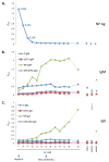
Nucleoprotein, virus-specific IgM and IgG detection by ELISA in G-1442, normal, and contact G-1446 sera. An Ag capture ELISA was used to detect LASV NP in patient sera (A). LASV NP Ag was not detected in normal sera from Sierra Leonean origin, or in contact G-1446. The level of LASV NP Ag [blue diamond] in G-1442 dropped significantly during the first 3 days of ribavirin administration, and was undetectable by day 10. LASV-specific IgM (B) and IgG (C) were assayed in a recombinant ELISA plate format, with individually coated NP, GP1 (sGP1), GP2 (GPCΔTM), or Z proteins. One Sierra Leonean serum registered a high IgG titer to NP (NHS015), whereas the other had moderate IgM titers to NP (NHS023), but both were negative for IgG and IgM to Z and glycoproteins. NP-specific IgM and IgG levels in G-1442 rose throughout the course of the illness, through day 20. Contact G-1446 did not have measurable IgG titers, and only registered a low IgM titer to Z. Data are plotted as mean A450 ± SD, N = -2. The line between day 18 and 20 is dotted to reflect discontinuity on day 19.
Figure 3
Figure 3
Comparison of LASV NP antigen detection by ELISA versus RNA quantification by qPCR. RNA was prepared from serum samples as outlined in materials and methods. RT-PCR followed by qPCR directed against the GPC gene was performed on days 7-18. A 1:6 dilution series of Josiah strain seed stock was used as a standard to calculate the LASV RNA copy number per milliliter of serum. PCR data were plotted on the second Y axis (LASV RNA copies/mL). Error-bars represent the SEM of two independent experiments. NP Ag ELISA data was plotted on the first Y axis (A450) for trend comparison. Trend lines for NP Ag ELISA (power) and qPCR (exponential), and associated R2 values are indicated. The limit of detection for antigen by NP Ag ELISA was day 9 (blue arrow), and day 11 for qPCR (red arrow).
Figure 4
Figure 4
Comprehensive daily Piccolo metabolic panel analysis. Metabolic indicators were measured in the serum of G-1442 daily after admission, through day 20 (with the exception of day 19), using a Piccolo comprehensive metabolic panel disk array. Two Sierra Leonean normal controls were also analyzed for comparison (SLN004 and SLN022). Values were plotted alongside normal ranges for each metabolite (rose boxes), for reference. G-1442 presented with normal BUN, CRE, but elevated TBIL. Analytes ALP, ALT, and AST were highly elevated upon admission, all indicative of severe liver implication in this LF case. Metabolic indicators in the two healthy Sierra Leonean donors were within normal ranges. Hemoglobin levels were independently measured in G-1442 on days 7, 9-12, and 14-16 post-onset of disease. The day of stillbirth delivery is indicated in each panel by a dark red diamond (day 13).
Figure 5
Figure 5
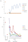
Serum cytokine levels analyzed by multiplex Flow Cytometry. Data generated with a Human 11-Plex Inflammatory Cytokine kit was quantified with Flow Cytomix Pro software, and plotted on linear scales. G-1442 presented with elevated levels of TNF-β(A.), IL-6, IL-8, IL-10, and notably IFN-γ(B.), all of which decreased by the next day, following initial treatment with ribavirin (day 7, arrow). Cytokine levels were also measured in normal Sierra Leonean controls, for comparison (LS004, LS022). Significant changes in the cytokine profile were not noted following delivery of a stillbirth fetus (day 13, arrow).
Figure 6
Figure 6
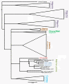
Phylogenetic analysis of LASV from patient G-1442. A ca. 800 bp fragment of the GPC gene was sequenced and aligned to 73 other sequences available in the NCBI database. A Neighbor-joining tree using LASV Pinneo as an outgroup was created with 1,000 replicates of bootstrap and clades from different countries are displayed as cartoons. Recent LASV isolates from the Kenema area are marked in blue. The scale bar indicates 3% nucleotide divergence.

References

    1. Buckley SM, Casals J. Lassa fever, a new virus disease of man from West Africa. Isolation and characterization of the virus. Am J Trop Med Hyg. 1970;19(4):680–691. - PubMed
    1. Birmingham K, Kenyon G. Lassa fever is unheralded problem in West Africa. Nat Med. 2001;7(8):878. - PubMed
    1. Fisher-Hoch SP, McCormick JB. Lassa fever vaccine: A review. Expert Rev Vaccines. 2004;3:103–111. - PubMed
    1. McCormick JB, King IJ, Webb PA, Johnson KM, O'Sullivan R, Smith ES, Trippel S, Tong TC, Sacchi N. A case-control study of the clinical diagnosis and course of Lassa fever. J Infect Dis. 1987;155(3):445–455. doi: 10.1093/infdis/155.3.445. - DOI - PubMed
    1. McCormick JB. Epidemiology and control of Lassa fever. Current Topics in Microbiol and Immunol. 1987;134:69–78. - PubMed

Publication types

MeSH terms