

Molecular adaptation of photoprotection: triplet states in light-harvesting proteins
- PMID: 21843485
- PMCID: PMC3175079
- DOI: 10.1016/j.bpj.2011.05.057
Molecular adaptation of photoprotection: triplet states in light-harvesting proteins
Abstract
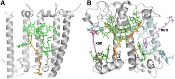
The photosynthetic light-harvesting systems of purple bacteria and plants both utilize specific carotenoids as quenchers of the harmful (bacterio)chlorophyll triplet states via triplet-triplet energy transfer. Here, we explore how the binding of carotenoids to the different types of light-harvesting proteins found in plants and purple bacteria provides adaptation in this vital photoprotective function. We show that the creation of the carotenoid triplet states in the light-harvesting complexes may occur without detectable conformational changes, in contrast to that found for carotenoids in solution. However, in plant light-harvesting complexes, the triplet wavefunction is shared between the carotenoids and their adjacent chlorophylls. This is not observed for the antenna proteins of purple bacteria, where the triplet is virtually fully located on the carotenoid molecule. These results explain the faster triplet-triplet transfer times in plant light-harvesting complexes. We show that this molecular mechanism, which spreads the location of the triplet wavefunction through the pigments of plant light-harvesting complexes, results in the absence of any detectable chlorophyll triplet in these complexes upon excitation, and we propose that it emerged as a photoprotective adaptation during the evolution of oxygenic photosynthesis.
Copyright © 2011 Biophysical Society. Published by Elsevier Inc. All rights reserved.
Figures
Similar articles
-
A photosynthetic antenna complex foregoes unity carotenoid-to-bacteriochlorophyll energy transfer efficiency to ensure photoprotection.Proc Natl Acad Sci U S A. 2020 Mar 24;117(12):6502-6508. doi: 10.1073/pnas.1920923117. Epub 2020 Mar 5. Proc Natl Acad Sci U S A. 2020. PMID: 32139606 Free PMC article.
-
Carotenoids and Photosynthesis.Subcell Biochem. 2016;79:111-39. doi: 10.1007/978-3-319-39126-7_4. Subcell Biochem. 2016. PMID: 27485220 Review.
-
Redox functions of carotenoids in photosynthesis.Biochemistry. 2004 Jul 13;43(27):8607-15. doi: 10.1021/bi0492096. Biochemistry. 2004. PMID: 15236568
-
Direct observation of triplet energy transfer between chlorophylls and carotenoids in the core antenna of photosystem I from Thermosynechococcus elongatus.Biochim Biophys Acta Bioenerg. 2024 Jan 1;1865(1):149016. doi: 10.1016/j.bbabio.2023.149016. Epub 2023 Oct 11. Biochim Biophys Acta Bioenerg. 2024. PMID: 37832862
-
Photosynthetic Light-Harvesting (Antenna) Complexes-Structures and Functions.Molecules. 2021 Jun 3;26(11):3378. doi: 10.3390/molecules26113378. Molecules. 2021. PMID: 34204994 Free PMC article. Review.
Cited by
-
Structure, regulation and assembly of the photosynthetic electron transport chain.Nat Rev Mol Cell Biol. 2025 Sep;26(9):667-690. doi: 10.1038/s41580-025-00847-y. Epub 2025 May 21. Nat Rev Mol Cell Biol. 2025. PMID: 40399647 Review.
-
Triplet-driven chemical reactivity of β-carotene and its biological implications.Nat Commun. 2022 May 5;13(1):2474. doi: 10.1038/s41467-022-30095-z. Nat Commun. 2022. PMID: 35513374 Free PMC article.
-
Excitation transfer and quenching in photosystem II, enlightened by carotenoid triplet state in leaves.Photosynth Res. 2024 Apr;160(1):31-44. doi: 10.1007/s11120-024-01086-6. Epub 2024 Mar 19. Photosynth Res. 2024. PMID: 38502255
-
Triplet-triplet energy transfer in artificial and natural photosynthetic antennas.Proc Natl Acad Sci U S A. 2017 Jul 11;114(28):E5513-E5521. doi: 10.1073/pnas.1614857114. Epub 2017 Jun 26. Proc Natl Acad Sci U S A. 2017. PMID: 28652359 Free PMC article.
-
A Hidden State in Light-Harvesting Complex II Revealed By Multipulse Spectroscopy.J Phys Chem B. 2015 Apr 23;119(16):5184-93. doi: 10.1021/acs.jpcb.5b01335. Epub 2015 Apr 10. J Phys Chem B. 2015. PMID: 25815531 Free PMC article.
References
-
- Truscott T.G., Land E.J., Sykes A. The in vitro photochemistry of biological molecules. 3. Absorption spectra, lifetimes and rates of oxygen quenching of the triplet states of β-carotene, retinal and related polyenes. Photochem. Photobiol. 1973;17:43–51. - PubMed
-
- Palozza P., Krinsky N.I. Antioxidant effects of carotenoids in vivo and in vitro: an overview. Methods Enzymol. 1992;213:403–420. - PubMed
-
- Pascal A.A., Liu Z., Ruban A. Molecular basis of photoprotection and control of photosynthetic light-harvesting. Nature. 2005;436:134–137. - PubMed
-
- Ruban A.V., Berera R., van Grondelle R. Identification of a mechanism of photoprotective energy dissipation in higher plants. Nature. 2007;450:575–578. - PubMed
Publication types
MeSH terms
Substances
LinkOut - more resources
Full Text Sources
