. 2011 Nov;176(2):143-50.
doi: 10.1016/j.jsb.2011.08.001. Epub 2011 Aug 6.

The effect of context on the folding of β-hairpins

Affiliations

The effect of context on the folding of β-hairpins

Amanda L Jonsson et al. J Struct Biol. 2011 Nov.

Abstract

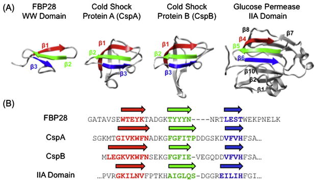
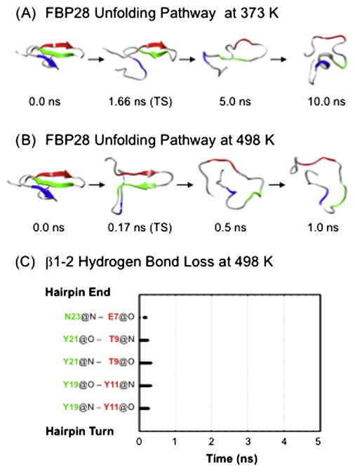
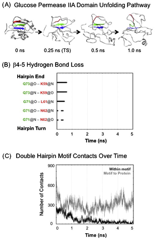
Small β-hairpin peptides have been widely used as models for the folding of β-sheets. But how applicable is the folding of such models to β-structure in larger proteins with conventional hydrophobic cores? Here we present multiple unfolding simulations of three such proteins that contain the WW domain double hairpin β-sheet motif: cold shock protein A (CspA), cold shock protein B (CspB) and glucose permease IIA domain. We compare the behavior of the free motif in solution and in the context of proteins of different size and architecture. Both Csp proteins lost contacts between the double-hairpin motif and the protein core as the first step of unfolding and proceeded to unfold with loss of the third β-strand, similar to the isolated WW domain. The glucose permease IIA domain is a larger protein and the contacts between the motif and the core were not lost as quickly. Instead the unfolding pathway of glucose permease IIA followed a different pathway with β1 pulling away from the sheet first. Interestingly, when the double hairpin motif was excised from the glucose permease IIA domain and simulated in isolation in water it unfolded by the same pathway as the WW domain, indicating that it is tertiary interactions with the protein that alter the motif's unfolding not a sequence dependent effect on its intrinsic unfolding behavior. With respect to the unfolding of the hairpins, there was no consistent order to the loss of hydrogen bonds between the β-strands in the hairpins in any of the systems. Our results show that while the folding behavior of the isolated WW domain is generally consistent with the double hairpin motif's behavior in the cold shock proteins, it is not the case for the glucose permease IIA domain. So, one must be cautious in extrapolating findings from model systems to larger more complicated proteins where tertiary interactions can overwhelm intrinsic behavior.


Figures

Fig. 1
Fig. 1. Double hairipin motif
A) Structures of FPB28 WW domain, CspA, CspB and glucose permease IIA domain with the double hairpin motif colored in red, green and blue. The extra strands in the β-sheet of glucose permease IIA domain are also labeled. For FBP28 WW domain residues 8–12 are red, 18–22 green, and 26–29 are blue for β1, β2 and β3, respectively. CspA is colored red for β1 residues 5–13, green for β2 residues 18–23, and blue for β3 residues 30–33. For CspB residues 2–10 are red for β1, 15–19 green for β2 and 26–29 blue for β3. Glucose permease IIA domain is colored red for β4 residues 58–63, green for β5 residues 69–74, and blue for β6 residues 79–83. The same coloring is used throughout the figures. B) Sequence alignment of the double hairpin motif in each system, with arrows marking the β-strands.
Fig. 2
Fig. 2. WW Domain Unfolding Pathway
A) Snapshots from a 373 K, low pH unfolding simulation. B) Snapshots from a 498 K, neutral pH unfolding simulation. C) The presence of hydrogen bonds in the first β-hairpin over time in the same simulation as shown in B. If a hydrogen bond is present between the specified atoms, a dot is graphed.
Fig. 3
Fig. 3. Cold Shock Protein Unfolding Pathway
A) Snapshots from simulation #1 of the CspA 498 K unfolding simulation. B) Hydrogen bonds between strands β1 and β2 over time for the simulation shown in panel A. C) Total number of atom-atom contacts over time for the simulation in panel A. In gray are contacts between the residues in the double hairpin motif and the rest of the protein. In black are contacts between just the residues in the double hairpin motif (strands β1–3). D) Snapshots from simulation #1 498 K CspB unfolding simulation.
Fig. 4
Fig. 4. Comparison of the experimental Φ-values and calculated S-values for the TS CspB
A representative structure from the TS ensemble is shown in Fig. 3D. The S-values were calculated over all structures in the TS ensemble pooled from all simulations. Points cover the 14 mutant set described in the text.
Fig. 5
Fig. 5. Glucose Permease IIA Domain Unfolding Pathway
A) Snapshots from simulation #1 of the IIA domain 498 K unfolding simulation. Note that the TS structure is representative of the TS ensemble, which has an average Cα RMSD within and between different simulations of 5.8 ± 0.8 Å. B) Hydrogen bonds between strands β4 and β5 over time for the simulation shown in panel A. C) Total number of atom-atom contacts over time for the simulation in panel A. In gray are contacts between the residues in the double hairpin motif and the rest of the protein. In black are contacts between just the residues in the double hairpin motif (strands β4–6).
Fig. 6
Fig. 6. Comparison of the Thermal Unfolding of the Double Hairpin Motif in Glucose Permease IIA Domain with the Excised Motif
A) The unfolding of the double hairpin motif in the context of the full IIA domain with the main side chains of residues involved in perturbing the motif’s unfolding pathway displayed and labeled. B) The unfolding of the excised double hairpin motif from the IIA domain. Note the similarities between the unfolding of this domain and the WW domain in Figure 2.
Fig. 7
Fig. 7. Comparison of the Thermal Unfolding of the isolate Double Hairpin Motif
A) FBP28 WW domain. B) Motif excised from CspA. C) Motif excised from CspB. D) Motif excised from glucose permease IIA domain. All simulations are at 498 K. Note the similarities in the unfolding of the secondary structure across all isolated domains despite differences in their sequences.

References

    1. Alonso DOV, DeArmond S, Cohen FE, Daggett V. Mapping the early steps of the pH-induced conformational conversion of the prion protein. Proc Natl Acad Sci USA. 2001;98:2985–2989. - PMC - PubMed
    1. Beck DAC, Alonso DOV, Daggett V. in lucem Molecular Mechanics (ilmm) University of Washington; Seattle: 2000–2011.
    1. Beck DAC, Daggett V. Methods for molecular dynamics simulations of protein folding/unfolding in solution. Methods Enzymol. 2004;34:112–120. - PubMed
    1. Beck DAC, Armen RS, Daggett V. Cutoff size need not strongly influence molecular dynamics results for solvated polypeptides. Biochemistry. 2005;44:609–616. - PubMed
    1. Beck DAC, Jonsson AL, Schaeffer RD, Scott KA, Day R, et al. Dynameomics: Mass annotation of protein dynamics and unfolding in water by high-throughput atomistic molecular dynamics simulations. Protein Eng, Des Sel. 2008;21:353–368. - PubMed

Publication types

MeSH terms

Substances

LinkOut - more resources