. 2011 Aug 29;208(9):1749-56.
doi: 10.1084/jem.20110578. Epub 2011 Aug 15.

Loss of Roquin induces early death and immune deregulation but not autoimmunity

Affiliations

Loss of Roquin induces early death and immune deregulation but not autoimmunity

Arianna Bertossi et al. J Exp Med. .

Abstract

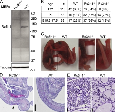
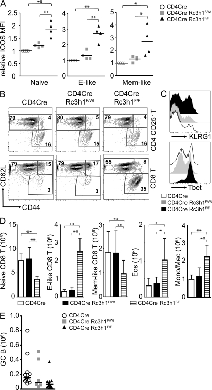
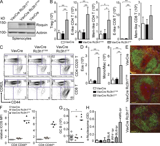
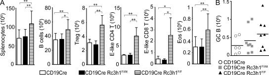
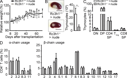
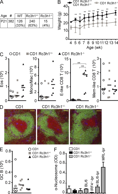
The substitution of one amino acid in the Roquin protein by the sanroque mutation induces a dramatic autoimmune syndrome in mice. This is believed to occur through ectopic expression of inducible T cell co-stimulator (ICOS) and unrestrained differentiation of follicular T helper cells, which induce spontaneous germinal center reactions to self-antigens. In this study, we demonstrate that tissue-specific ablation of Roquin in T or B cells, in the entire hematopoietic system, or in epithelial cells of transplanted thymi did not cause autoimmunity. Loss of Roquin induced elevated expression of ICOS through T cell-intrinsic and -extrinsic mechanisms, which itself was not sufficient to break self-tolerance. Instead, ablation of Roquin in the hematopoietic system caused defined changes in immune homeostasis, including the expansion of macrophages, eosinophils, and T cell subsets, most dramatically CD8 effector-like T cells, through cell-autonomous and nonautonomous mechanisms. Germline Roquin deficiency led to perinatal lethality, which was partially rescued on the genetic background of an outbred strain. However, not even complete absence of Roquin resulted in overt self-reactivity, suggesting that the sanroque mutation induces autoimmunity through an as yet unknown mechanism.


Figures

Figure 1.
Figure 1.
Loss of Roquin causes perinatal lethality. (A) Western blot of Roquin protein expression in wild-type and Rc3h1−/− immortalized murine embryonic fibroblasts. (B) Genotype frequency of offspring from intercrosses of Rc3h1+/− mice. (C) Newborn (P0) Rc3h1−/− pups show a curly tail. (D) PAS stainings of sagittal sections of the sacral spinal column of Rc3h1−/− and control newborn mice; the arrow indicates the skin, soft tissue, and bone defect. (E) PAS stainings of sagittal sections of lungs of Rc3h1−/− and control newborn mice. Bars: (D) 1 mm; (E) 50 µm.
Figure 2.
Figure 2.
Roquin deficiency in T lymphocytes leads to CD8 effector–like T cell expansion. (A) Relative MFI of ICOS expression on CD4 T cell subsets in the spleens of mice of the indicated genotypes. Naive, CD25CD44loCD62Lhi; effector (E)-like, CD25CD44hiCD62Llo; memory (Mem)-like, CD25CD44hiCD62Lhi. Bars indicate medians of four mice per group, and ICOS levels on CD4cre control T cell subsets are set to 1. (B) Representative contour plots of T cell (TCR-β+) subsets gated as indicated on the right. Numbers represent percentages of the gated populations of the indicated T cell subtype. (C) Histogram of KLRG1 and Tbet expression on CD44hiCD62Llo-gated effector-like CD8 T cells, representative of five experiments. (D) Bar charts of absolute numbers of indicated T cell (TCR-β+; subsets as in A) and myeloid cell subsets: Eos, eosinophils (Gr1intSiglecF+); Mono/Mac, monocytes/macrophages (SiglecFGr1intMac1+). Columns and error bars indicate means ± SD calculated from six to nine mice per genotype. (E) Absolute numbers of splenic germinal center (GC; B220+PNAhiFashiCD38lo) B cells of 11–6 mice per genotype. Bars indicate the median. *, P < 0.05; **, P < 0.001; one-way ANOVA.
Figure 3.
Figure 3.
Deregulation of immune homeostasis in the absence of Roquin in the hematopoietic system. (A) Western blot analysis to evaluate Roquin protein expression in splenocytes. (B and C) Absolute cell numbers (B) and representative contour plots (C) of splenic T cell (TCR-β+) subsets: effector (E)-like, CD44hiCD62Llo; and memory (Mem)-like, CD44hiCD62Lhi (n = 7–9); Treg cells, CD4+FoxP3+ (n = 3–5). Columns and error bars represent means ± SD calculated from the number of mice indicated. (D) Columns and error bars represent means ± SD of absolute cell numbers of eosinophils (Eos; Gr1intSiglecF+) and monocytic/macrophages (Mono/Mac; SiglecFGr1intMac1+) calculated from four to six mice per group. (E) Immunofluorescence of spleen sections: green, α-B220; red, α-CD3; blue, α-laminin. Bars, 100 µm. (F) Relative ICOS MFI of naive (CD44lo) and memory/effector-like (CD44hi) splenic CD4 T cells. Bars indicate medians. (G) Absolute cell numbers of germinal center (GC; B220+PNAhiFashiCD38lo) B cells. Bars represent medians calculated from five to seven mice per genotype. (H) Titers of serum autoantibodies against nucleosomes measured by ELISA (2–7 mo old) from 8–10 mice of the indicated genotypes. Sera from BL/6, IRF4−/−-lpr, BL/6-lpr, and MRL-lpr mice were used as positive or negative controls. Columns and error bars represent means ± SD. *, P < 0.05; **, P < 0.001; one-way ANOVA.
Figure 4.
Figure 4.
Roquin-deficient B cells cause effector-like T cell expansion. (A) Column and error bars represent means ± SD of absolute cell numbers of splenocytes (n = 8), B cells (B220+; n = 8), Treg cells (CD4+FoxP3+; n = 4), CD4 effector (E)–like T cells (CD25CD44hiCD62Llo; n = 4), CD8 effector–like T cells (CD44hiCD62Llo; n = 4), and eosinophils (Eos; Gr1intSiglecF+; n = 8). (B) Absolute cell numbers of germinal center (GC; B220+FashiPNAhiCD38lo) B cells. Bars indicate medians calculated from six to eight mice per group. *, P < 0.05; **, P < 0.001; one-way ANOVA.
Figure 5.
Figure 5.
Effect of Roquin loss in thymic epithelial cells. (A) Growth curve of nude mice transplanted with fetal thymic epithelium as indicated. Means ± SD for recipients of six wild-type and six Rc3h1−/− thymi are shown. (B) Rc3h1+/+ and Rc3h1−/− thymic epithelium–derived thymus lobes on kidneys of C57BL/6 nude mice 10 wk after transplantation. Bar chart shows absolute numbers of thymocytes. Bars, 500 µm. (C) Proportions of thymocyte subsets. DN, (CD4/CD8) double negative; DP, double positive; SP, single positive; Treg cells, CD4+FoxP3+. (D) Usage of individual Vα (left) and Vβ (right) chains in the TCR on CD4 T cells in mice of the indicated genotypes. (B–D) Columns and error bars represent means ± SD of five to six mice per group. **, P < 0.001; Student’s t test.
Figure 6.
Figure 6.
Roquin knockout mice can survive on a CD1 outbred genetic background. (A) Genotype frequency of offspring of heterozygous matings of CD1 Rc3h1+/− mice. (B) Body weight curve of Rc3h1−/− mice compared with control littermates (2–21 mice per group and time point). Error bars indicate SD. (C and E) Absolute cell numbers of eosinophils (Eos; Gr1intSiglec-F+), monocytic/macrophages (Mono/Mac; SiglecFGr1intMac1+), splenic CD8+ T cell subsets (Effector [E]-like, CD44hiCD62Llo; memory (Mem)-like, CD44hiCD62Lhi; C) and germinal center (GC; B220+PNAhiFashiCD38lo) B cells (E) calculated from six mice of the indicated genotypes. Bars represent medians. (D) Immunofluorescence of spleen sections: green, α-B220; red, α-CD3; blue, α-laminin. Bars, 100 µm. (F) Titers of autoantibodies against nucleosomes detected by ELISA in the serum of eight to nine 2.5–3.5-mo-old mice of the indicated genotypes. Columns and error bars indicate means ± SD. **, P < 0.001; one-way ANOVA.

References

    1. Athanasopoulos V., Barker A., Yu D., Tan A.H., Srivastava M., Contreras N., Wang J., Lam K.P., Brown S.H., Goodnow C.C., et al. 2010. The ROQUIN family of proteins localizes to stress granules via the ROQ domain and binds target mRNAs. FEBS J. 277:2109–2127 10.1111/j.1742-4658.2010.07628.x - DOI - PubMed
    1. Betz U.A., Vosshenrich C.A., Rajewsky K., Müller W. 1996. Bypass of lethality with mosaic mice generated by Cre-loxP-mediated recombination. Curr. Biol. 6:1307–1316 10.1016/S0960-9822(02)70717-3 - DOI - PubMed
    1. de Boer J., Williams A., Skavdis G., Harker N., Coles M., Tolaini M., Norton T., Williams K., Roderick K., Potocnik A.J., Kioussis D. 2003. Transgenic mice with hematopoietic and lymphoid specific expression of Cre. Eur. J. Immunol. 33:314–325 10.1002/immu.200310005 - DOI - PubMed
    1. Glasmacher E., Hoefig K.P., Vogel K.U., Rath N., Du L., Wolf C., Kremmer E., Wang X., Heissmeyer V. 2010. Roquin binds inducible costimulator mRNA and effectors of mRNA decay to induce microRNA-independent post-transcriptional repression. Nat. Immunol. 11:725–733 10.1038/ni.1902 - DOI - PubMed
    1. Goodnow C.C. 2007. Multistep pathogenesis of autoimmune disease. Cell. 130:25–35 10.1016/j.cell.2007.06.033 - DOI - PubMed

Publication types

MeSH terms

Substances