. 2011 Aug 29;208(9):1757-65.
doi: 10.1084/jem.20102170. Epub 2011 Aug 15.

The Paf oncogene is essential for hematopoietic stem cell function and development

Affiliations

The Paf oncogene is essential for hematopoietic stem cell function and development

Yacine M Amrani et al. J Exp Med. .

Abstract

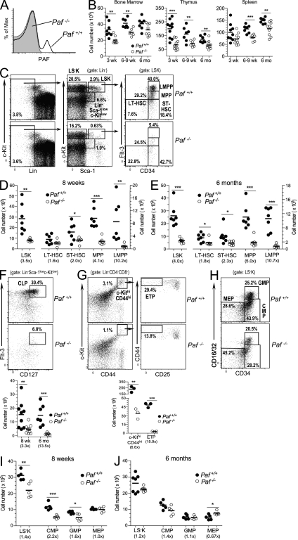
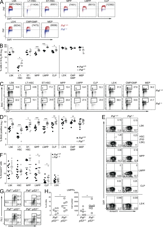
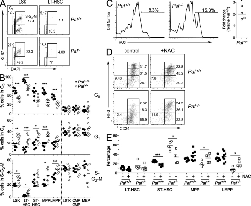
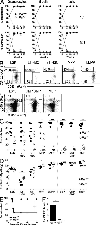
Hematopoietic stem cells (HSCs) self-renew to maintain the lifelong production of all blood populations. Here, we show that the proliferating cell nuclear antigen-associated factor (Paf) is highly expressed in cycling bone marrow HSCs and plays a critical role in hematopoiesis. Mice lacking Paf exhibited reduced bone marrow cellularity; reduced numbers of HSCs and committed progenitors; and leukopenia. These phenotypes are caused by a cell-intrinsic blockage in the development of long-term (LT)-HSCs into multipotent progenitors and preferential loss of lymphoid progenitors caused by markedly increased p53-mediated apoptosis. In addition, LT-HSCs from Paf(-/-) mice had increased levels of reactive oxygen species (ROS), failed to maintain quiescence, and were unable to support LT hematopoiesis. The loss of lymphoid progenitors was likely due the increased levels of ROS in LT-HSCs caused by treatment of Paf(-/-) mice with the anti-oxidant N-acetylcysteine restored lymphoid progenitor numbers to that of Paf(+/+) mice. Collectively, our studies identify Paf as a novel and essential regulator of early hematopoiesis.


Figures

Figure 1.
Figure 1.
Paf is necessary for proper HSC and progenitor development. (A) FACS of thymocytes from Paf+/+ and Paf−/− mice stained for intracellular PAF. (B) Total number of cells in BM, thymuses, and spleens of Paf+/+ and Paf−/− mice. (C) Representative staining profiles for BM HSC and progenitor populations. (D and E) Absolute cell numbers in the BM of 8-wk-old (D) or 6-mo-old (E) Paf+/+ and Paf−/− mice. (F) Representative staining profiles and absolute number of CLPs in the BM of 8-wk- and 6-mo-old mice. Plots are gated for LinSca-1lowc-kitlow cells as defined in (C). (G) Representative staining profiles and absolute cell numbers of ETPs in the thymus of 8-wk-old mice. (H) Representative staining profiles for myeloerythroid progenitors. LSK cells (defined in C) were subdivided into CMPs, GMPs, and MEPs as indicated. (I and J). Absolute cell numbers of LSKs, CMPs, GMPs, and MEPs in the BM of 8-wk-old (I) or 6-mo-old (J) mice. Each symbol represents a mouse combined from 3–6 experiments. Horizontal bars indicate the mean. Numbers in parentheses are the fold change of cell numbers for Paf−/− relative to Paf+/+ mice. *, P < 0.05; **, P < 0.01; ***, P < 0.001.
Figure 2.
Figure 2.
Paf restricts HSC and progenitor cycling and protects LMPPs and CLPs from p53-mediated apoptosis. (A) PAF protein levels in BM HSC and progenitor populations relative to cell cycle (DAPI). Data are presented as an overlay of PAF staining obtained for each population from Paf+/+ and Paf−/− mice. Numbers in parentheses are the MFI obtained for PAF. Horizontal light blue line highlights the differences in PAF expression among the populations. LSK cells were separated into MEPs (CD34) and a population containing CMPs and GMPs (CD34+). (B) Percentage of BM Paf+/+ and Paf−/− HSC and progenitors that are in the S–G2–M phases of the cell cycle. Representative FACS profiles are shown in Fig. S2 D. (C) Representative staining profiles of Paf+/+ and Paf−/− BM cells from mice that were injected 24 h earlier with BrdU. The BrdU+ gate was established by using BM from PBS-injected mice (Fig. S2 E). (D) Percentage of each BM subset that incorporated BrdU. (E) Representative FACS analyses of HSC subsets showing Annexin V versus DAPI staining. Gating of apoptotic cells (AnnexinV+DAPI) was determined as shown in Fig. S2 F. (F) Cumulative data showing the percentage of cells undergoing apoptosis. (G) Representative staining profiles for LT-HSC, ST-HSC, MPP, and LMPP from Paf+/+p53+/+, Paf+/+p53+/−, Paf−/−p53+/+, and Paf−/−p53+/− mice. (H) Cumulative data showing the LMPP frequency (left) and absolute cell numbers (right). Data were combined from 3–8 experiments on 1–2 mice/genotype. Each symbol represents a mouse, and horizontal bars mark the mean. *, P < 0.05; **, P < 0.01; ***, P < 0.001.
Figure 3.
Figure 3.
Disruption of HSC quiescence in the absence of Paf. (A) Representative staining profiles for LSK and LT-HSC analyzed for Ki-67 and cell cycle (DAPI). (B) Cumulative data showing the percentage of cells in each BM subset that are quiescent (G0), resting (G1), or cycling (S–G2–M). (C) Representative histograms showing Lin BM LT-HSCs loaded with the ROS detection reagent, 5-(and-6)-carboxy-2’,7’-difluorodihydrofluorescein diacetate. The gate shows the percentage of ROS+ cells. Cumulative data shows the fold difference in percentage of ROS positive LT-HSCs for Paf−/− relative to Paf+/+ LT-HSCs. (D) Representative FACS profiles for BM subsets from NAC-treated or control mice. (E) Cumulative data showing the frequency of each population as a percentage of the LSK gate from (D). Absolute cell numbers are shown in Fig. S3 D. Data were combined from 3–4 individual experiments of 1–3 mice/genotype. Each symbol represents a mouse and horizontal bars indicate the mean. *, P < 0.05; **, P < 0.01; ***, P < 0.001.
Figure 4.
Figure 4.
Paf−/− HSCs are functionally compromised. (A) Peripheral blood from lethally irradiated mice transplanted with a 1:1 (n = 5) or 9:1 (n = 6) mix of Lin BM from Paf −/− (CD45.2) and WT (CD45.1) donor mice were monitored for the contribution of Paf +/+ and Paf −/− HSCs to the granulocyte, T and B cell lineages. Data show mean ± SD and are representative of two identical experiments. (B) Representative FACS analysis of BM HSC populations at 20 wk after transplant for 9:1 BM-transplanted mice. (C) Cumulative data from B. Data were pooled from analyses of three mice/genotype for two independent chimera experiments. (D) Cumulative data showing the percentage of cycling cells in the BM of 9:1 transplanted mice (representative FACS plots are shown in Fig. S3 F). Data pooled for two mice from two independent experiments. (E) Paf−/− BM cells are defective in rescuing lethally irradiated recipients after a second transplant. Survival curves for recipients of secondary BM transplants. Data combined from two independent experiments of 10 mice/genotype each. (F) The number of cobblestone area forming cells in Lin Paf+/+ and Paf−/− BM was determined by scoring colonies at 21 d. Data shown are means of two similar experiments ± SD of 1 mouse/genotype. *, P < 0.05; **, P < 0.01; ***, P < 0.001.

Similar articles

Cited by

References

    1. Adolfsson J., Borge O.J., Bryder D., Theilgaard-Mönch K., Astrand-Grundström I., Sitnicka E., Sasaki Y., Jacobsen S.E. 2001. Upregulation of Flt3 expression within the bone marrow Lin(-)Sca1(+)c-kit(+) stem cell compartment is accompanied by loss of self-renewal capacity. Immunity. 15:659–669 10.1016/S1074-7613(01)00220-5 - DOI - PubMed
    1. Akashi K., Traver D., Miyamoto T., Weissman I.L. 2000. A clonogenic common myeloid progenitor that gives rise to all myeloid lineages. Nature. 404:193–197 10.1038/35004599 - DOI - PubMed
    1. Akilesh S., Petkova S., Sproule T.J., Shaffer D.J., Christianson G.J., Roopenian D. 2004. The MHC class I-like Fc receptor promotes humorally mediated autoimmune disease. J. Clin. Invest. 113:1328–1333 - PMC - PubMed
    1. Breems D.A., Blokland E.A., Neben S., Ploemacher R.E. 1994. Frequency analysis of human primitive haematopoietic stem cell subsets using a cobblestone area forming cell assay. Leukemia. 8:1095–1104 - PubMed
    1. Chi A.W., Bell J.J., Zlotoff D.A., Bhandoola A. 2009. Untangling the T branch of the hematopoiesis tree. Curr. Opin. Immunol. 21:121–126 10.1016/j.coi.2009.01.012 - DOI - PMC - PubMed

Publication types

MeSH terms