. 2011 Nov;79(11):4657-67.
doi: 10.1128/IAI.05583-11. Epub 2011 Aug 15.

The novel leptospiral surface adhesin Lsa20 binds laminin and human plasminogen and is probably expressed during infection

Affiliations

The novel leptospiral surface adhesin Lsa20 binds laminin and human plasminogen and is probably expressed during infection

Renata Siqueira Mendes et al. Infect Immun. 2011 Nov.

Abstract

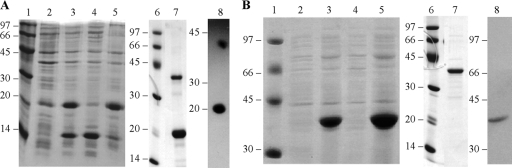
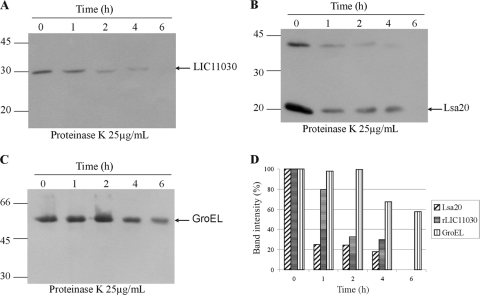
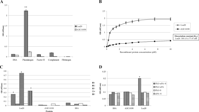
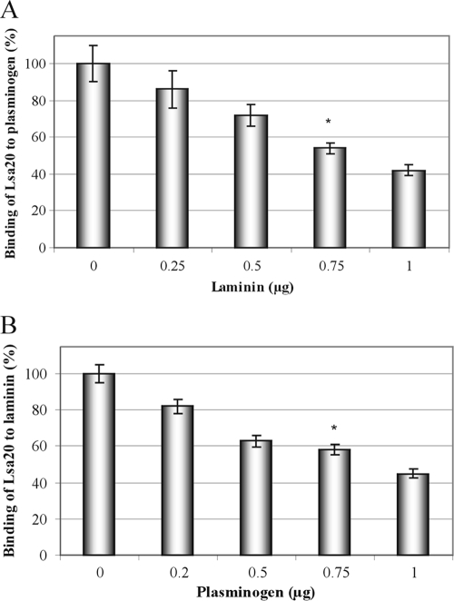
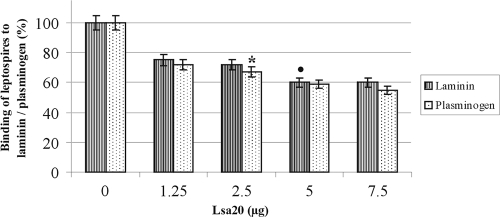
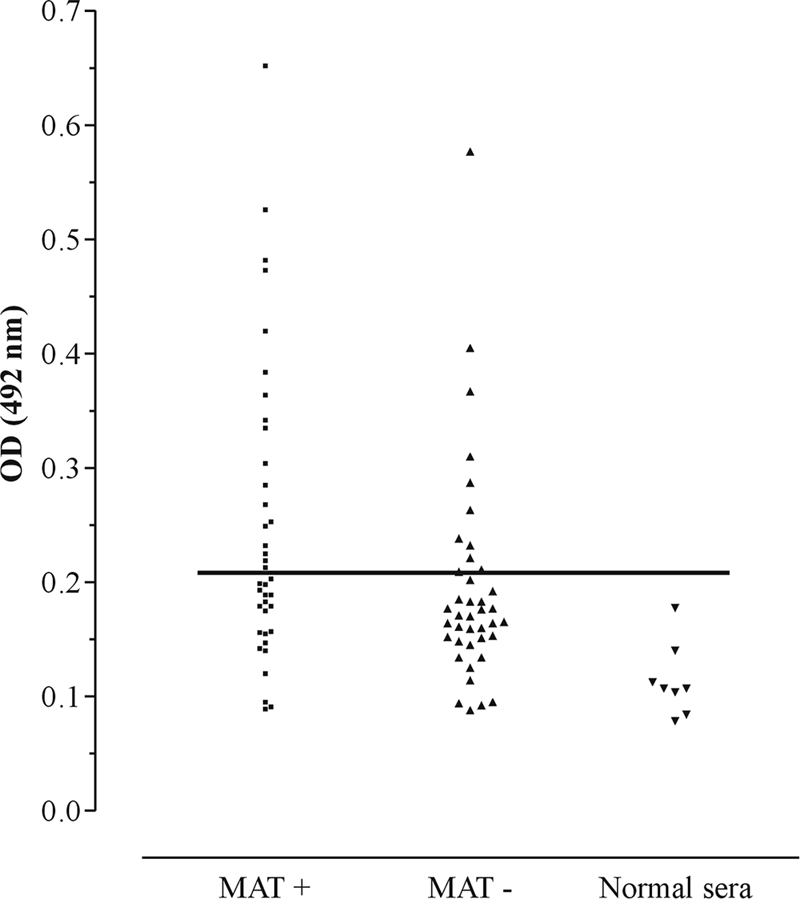
Leptospirosis is an emerging infectious disease caused by pathogenic species of Leptospira. In this work, we report the cloning, expression, purification, and characterization of two predicted leptospiral outer membrane proteins, LIC11469 and LIC11030. The LIC11469 protein is well conserved among leptospiral strains, while LIC11030 was identified only in Leptospira interrogans. We confirmed by surface proteolysis of intact leptospires with proteinase K that these proteins are most likely new surface leptospiral proteins. The recombinant proteins were evaluated for their capacity to attach to extracellular matrix (ECM) components and to plasminogen. The leptospiral protein encoded by LIC11469, named Lsa20 (leptospiral surface adhesin of 20 kDa), binds to laminin and to plasminogen. The binding with both components was not detected when Lsa20 was previously denatured or blocked with anti-Lsa20 antibodies. Moreover, Lsa20 binding to laminin was also confirmed by surface plasmon resonance (SPR). Laminin competes with plasminogen for binding to Lsa20, suggesting the same ligand-binding site. Lsa20-bound plasminogen could be converted to enzymatically active plasmin, capable of cleaving plasmin substrate d-valyl-leucyl-lysine-p-nitroanilide dihydrochloride. Lsa20 was recognized by antibodies in confirmed-leptospirosis serum samples, suggesting that this protein is expressed during infection. Taken together, our results indicate that Lsa20 is a novel leptospiral adhesin that in concert with the host-derived plasmin may help the bacteria to adhere and to spread through the hosts.


Figures

Fig. 1.
Fig. 1.
Recombinant protein analysis by SDS-PAGE and Western blotting. Lsa20 (A) and rLIC11030 (B) protein expression from NaCl-induced E. coli BL21-SI and IPTG-induced E. coli BL21(DE3) Star pLysS cultures, respectively. Lanes: 1 and 6, molecular mass protein marker; 2, noninduced total bacterial extract; 3, total bacterial cell lysates after induction; 4 and 5, soluble and insoluble (pellet) fractions of the induced culture, respectively; 7, purified recombinant proteins in Coomassie blue-stained gels; 8, Western blotting of the recombinant proteins probed with anti-His tag antibodies.
Fig. 2.
Fig. 2.
Conservation of LIC11469 and LIC11030 coding sequences among leptospira strains. Whole-cell lysates, Lsa20, and rLIC11030 were separated by SDS-PAGE, transferred into membranes, and probed with antiserum against each recombinant protein followed by anti-mouse IgG conjugated to peroxidase. Reactivity was detected by ECL kit and X-ray film exposure. Western blotting of gel transferred to membrane probed with anti-Lsa20 (A) and with anti-rLIC11030 (B). On the right are lanes (+) containing the respective recombinant proteins as markers.
Fig. 3.
Fig. 3.
Protease accessibility assay of LIC11469- and LIC11030-encoded proteins of L. interrogans. Viable leptospires were incubated with the indicated concentrations of proteinase K. Approximately 30 μg of total leptospiral extract was loaded per lane and separated by SDS-PAGE. After transfer, the immobilized extracts were subjected to immunoblot analysis using Lsa20 (A)- and rLIC11030 (B)-specific antibodies. (C) Antibodies specific for the protoplasmic cylinder protein GroEL were employed. The serum dilution was 1:100. The GroEL protein (same amount of cell extract) was used as an indicator of outer membrane integrity and as an internal control for proteinase K resistance. (D) Optical densities of the protein bands were taken, and the relative percentage was based on the corresponding protein amount at time zero (no proteinase K addition).
Fig. 4.
Fig. 4.
Binding characteristics of Lsa20 and rLIC11030 to ECM components. (A) Wells were coated with 1 μg of laminin, collagen type I, collagen type IV, cellular fibronectin, plasma fibronectin, ECM gel, and the control proteins BSA and fetuin. One microgram of the recombinant proteins was added per well, and the binding was measured by an ELISA. Data represent the means ± standard deviations of results from three independent experiments. For statistical analyses, the attachment of recombinant proteins to the ECM components was compared to its binding to BSA as well as to fetuin by the two-tailed t test (*, P < 0.005). (B) Lsa20 dose-dependent binding experiments: each point was performed in triplicate and is expressed as the mean absorbance value at 492 nm ± standard error for each point. Protein rLIC11030 was included as the negative control. (B) The dissociation constant (Kd) was calculated based on ELISA data for the Lsa20 recombinant protein, which reached equilibrium at a concentration of ∼9 μM. (C) Wells were coated with 1 μg of laminin, Lsa20, rLIC11030, or BSA (control). One microgram of the proteins was added per well, and the binding was measured by an ELISA. Bars: 1, coating with laminin followed by recombinant protein or BSA and incubation with anti-polyhistidine monoclonal antibodies; 2, coating with laminin followed by incubation with recombinant protein previously blocked by incubation with specific antibodies produced in hamster, and then incubation with anti-polyhistidine monoclonal antibodies; 3, coating with laminin followed by recombinant protein or BSA and incubation with specific mouse antirecombinant protein; 4, coating with laminin followed by incubation with denatured recombinant protein and incubation with specific mouse antirecombinant protein; 5, coating with recombinant protein or BSA followed by incubation with laminin and antilaminin antibodies. Data represent the means ± standard deviations of results from two independent experiments. For statistical analyses, the attachment of recombinant proteins to laminin was compared to its binding to BSA by the two-tailed t test (*, P < 0.02). (D) Analysis of the interaction of Lsa20 with laminin by using the BIAcore T100 system. Protein solutions of Lsa20 (0, 0.312, 0.625, 1.25, 2.5, 5.0, 10 μM) in 10 mM HEPES, 150 mM NaCl, 3 mM EDTA, 0.005% surfactant P20 (pH 7.4) were injected over immobilized laminin at a flow rate of 30 μl/min for 1 min at 25°C.
Fig. 5.
Fig. 5.
Recombinant protein Lsa20 binds human plasminogen and generates enzymatically active plasmin. (A) Components were coated into ELISA plates (10 μg/ml) and allowed to interact with 1 μg per well of the recombinant proteins Lsa20 and rLIC11030. The binding was detected by specific serum antirecombinant proteins. Bars represent the mean of absorbance at 492 nm ± standard deviation of three replicates for each protein and are representative of three independent experiments. For statistical analyses, the binding of Lsa20 to human plasminogen was compared to its binding to BSA by a two-tailed t test (**, P < 0.0005). (B) Plasminogen (10 μg/ml) was immobilized into 96-well ELISA plates, and 0 to 10 μM each recombinant protein was added for interaction. Protein rLIC11030 was included as the negative control. The binding was detected using antiserum raised in mice against the recombinant proteins in appropriate dilutions (1:1,000 for Lsa20) followed by horseradish peroxidase-conjugated anti-mouse IgG. Data represent the mean absorbance values ± standard deviation of three replicates for each experimental group. (B) The dissociation constant (Kd) was calculated based on ELISA data for Lsa20 recombinant protein when the equilibrium concentration was reached. (C) Wells were coated with 1 μg of PLG, Lsa20, rLIC11030, or BSA (control). One microgram of the proteins was added per well, and the binding was measured by an ELISA. Bars: 1, coating with PLG followed by recombinant protein or BSA and incubation with anti-polyhistidine monoclonal antibodies; 2, coating with PLG followed by incubation with recombinant protein previously blocked by incubation with specific antibodies produced in hamster, and then incubation with anti-polyhistidine monoclonal antibodies; 3, coating with PLG followed by recombinant protein or BSA and incubation with specific mouse antirecombinant protein; 4, coating with PLG followed by incubation with denatured recombinant protein and incubation with specific mouse antirecombinant protein; 5, coating with recombinant protein or BSA followed by incubation with PLG and anti-PLG antibody. Data represent the means ± standard deviations of results from two independent experiments. For statistical analyses, the attachment of recombinant proteins to laminin was compared to its binding to BSA by the two-tailed t test (*, P < 0.02). (D) Plasmin generation by plasminogen bound to recombinant protein Lsa20 was assayed by modified ELISA for immobilized proteins receiving treatment with PLG plus uPA plus specific plasmin substrate (PLG+uPA+S) or control treatments lacking one of the three components (PLG+uPA, PLG+S, uPA+S). BSA was employed as the negative control. Bars represent mean absorbance at 405 nm, as a measure of relative substrate degradation ± standard deviation of four replicates for each experimental group, and are representative of two independent experiments. Statistically significant binding in comparison to the negative control (BSA) is depicted: *, P < 0.003.
Fig. 6.
Fig. 6.
Laminin and plasminogen compete for the binding to Lsa20. (A) The effect of laminin on the binding of plasminogen (10 μg/ml) to immobilized Lsa20 (10 μg/ml) was assessed with the addition of increasing concentrations of laminin (0 to 1.00 μM). (B) The effect of plasminogen on the binding of laminin (10 μg/ml) to immobilized Lsa20 (10 μg/ml) was assessed with the addition of increasing concentrations of plasminogen (0 to 1.00 μM). The detection of Lsa20-bound plasminogen (A) or Lsa20-bound laminin (B) was performed by use of specific antibodies. Bars represent the mean absorbance values ± standard deviation of four replicates for each condition and are representative of two independent experiments. Results of statistically significant interference with binding in comparison with the positive control (no addition of laminin or plasminogen) are shown: *, P < 0.05.
Fig. 7.
Fig. 7.
Inhibition of L. interrogans attachment to laminin or to plasminogen by Lsa20. Laminin or plasminogen (1 μg/well) was adsorbed to microtiter plates followed by incubation with increasing concentrations of Lsa20 (0 to 7.50 μg) for 90 min at 37°C. Live leptospires (100 μl/well of 4 × 10⁷ L. interrogans serovar Copenhageni strain M20 leptospires) were added for 90 min at 37°C. The unbound leptospires were washed, and the quantification of bound leptospires was performed indirectly by anti-LipL32 antibodies produced in mice (1: 4,000 dilution) followed by horseradish peroxidase-conjugated anti-mouse IgG antibodies. Each point represents the mean absorbance value at 492 nm ± standard deviation of three replicates. Data are representative of two independent experiments. *, P < 0.009; •, P < 0.005.
Fig. 8.
Fig. 8.
Recognition of recombinant protein Lsa20 by IgG antibodies of individuals diagnosed with leptospirosis. Positive sera (responders) were determined by ELISA with the recombinant protein and serum samples from patients in both phases of the disease. The reactivity was evaluated as IgG antibodies. Serum was considered MAT positive or MAT negative if agglutination was detected when the sera were tested for their reactivities with isolates of the 22 serovars (see Materials and Methods). The cutoff values are defined as the mean plus 3 standard deviations obtained for sera from five healthy individuals.

Similar articles

Cited by

References

    1. Adu-Bobie J., Capecchi B., Serruto D., Rappuoli R., Pizza M. 2003. Two years into reverse vaccinology. Vaccine 21:605–610 - PubMed
    1. Altschul S. F., Gish W., Miller W., Myers E. W., Lipman D. J. 1990. Basic local alignment search tool. J. Mol. Biol. 215:403–410 - PubMed
    1. Angles-Cano E. 1994. Overview on fibrinolysis: plasminogen activation pathways on fibrin and cell surfaces. Chem. Phys. Lipids 67-68:353–362 - PubMed
    1. Angles-Cano E., de la Pena Diaz A., Loyau S. 2001. Inhibition of fibrinolysis by lipoprotein(a). Ann. N. Y. Acad. Sci. 936:261–275 - PubMed
    1. Atzingen M. V., et al. 2008. Lsa21, a novel leptospiral protein binding adhesive matrix molecules and present during human infection. BMC Microbiol. 8:70. - PMC - PubMed

Publication types

Associated data

LinkOut - more resources