. 2011 Nov;79(11):4696-707.
doi: 10.1128/IAI.05658-11. Epub 2011 Aug 15.

Proteomic analysis of Anaplasma phagocytophilum during infection of human myeloid cells identifies a protein that is pronouncedly upregulated on the infectious dense-cored cell

Affiliations

Proteomic analysis of Anaplasma phagocytophilum during infection of human myeloid cells identifies a protein that is pronouncedly upregulated on the infectious dense-cored cell

Matthew J Troese et al. Infect Immun. 2011 Nov.

Abstract

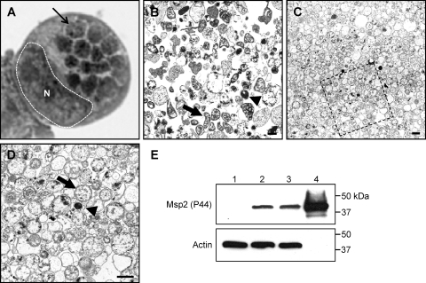
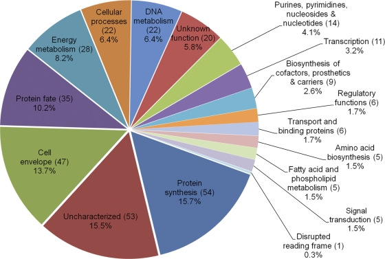
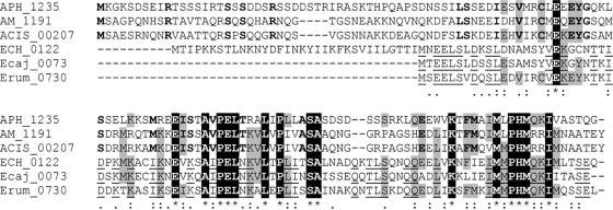
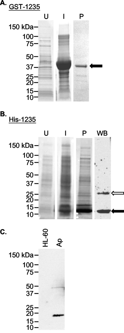
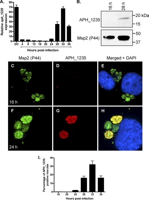
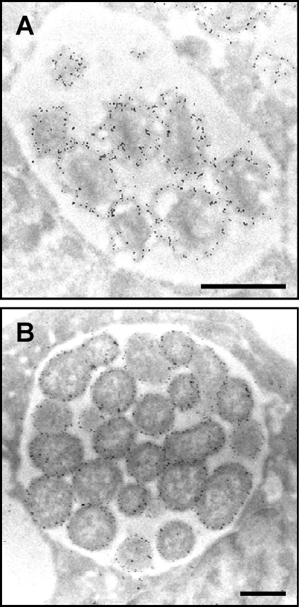
Anaplasma phagocytophilum is an obligate intracellular bacterium that invades neutrophils to cause the emerging infectious disease human granulocytic anaplasmosis. A. phagocytophilum undergoes a biphasic developmental cycle, transitioning between an infectious dense-cored cell (DC) and a noninfectious reticulate cell (RC). To gain insights into the organism's biology and pathogenesis during human myeloid cell infection, we conducted proteomic analyses on A. phagocytophilum organisms purified from HL-60 cells. A total of 324 proteins were unambiguously identified, thereby verifying 23.7% of the predicted A. phagocytophilum proteome. Fifty-three identified proteins had been previously annotated as hypothetical or conserved hypothetical. The second most abundant gene product, after the well-studied major surface protein 2 (P44), was the hitherto hypothetical protein APH_1235. APH_1235 homologs are found in other Anaplasma and Ehrlichia species but not in other bacteria. The aph_1235 RNA level is increased 70-fold in the DC form relative to that in the RC form. Transcriptional upregulation of and our ability to detect APH_1235 correlate with RC to DC transition, DC exit from host cells, and subsequent DC binding and entry during the next round of infection. Immunoelectron microscopy pronouncedly detects APH_1235 on DC organisms, while detection on RC bacteria minimally, at best, exceeds background. This work represents an extensive study of the A. phagocytophilum proteome, discerns the complement of proteins that is generated during survival within human myeloid cells, and identifies APH_1235 as the first known protein that is pronouncedly upregulated on the infectious DC form.


Figures

Fig. 1.
Fig. 1.
Dounce homogenization followed by discontinuous gradient centrifugation purifies A. phagocytophilum organisms while minimizing host cellular material contamination. (A) Light microscopic image of a Hema Fix-stained A. phagocytophilum-infected HL-60 cell that exhibits an infection load that is typical of the infected cells from which A. phagocytophilum organisms were purified for this study. A hatched line and an N demarcate the host cell nucleus, while a thin arrow denotes one of 14 morulae. (B) Transmission electron micrograph of A. phagocytophilum organisms and HL-60 cellular debris following syringe lysis and differential centrifugation to partially remove host cellular material. (C and D) Transmission electron micrograph of an A. phagocytophilum fraction obtained following centrifugation of a Dounce homogenate through a discontinuous Optiprep gradient. (D) Magnification of the region in panel C that is denoted by a hatched box. Thick arrows and arrowheads denote representative RC and DC organisms, respectively. Scale bars, 1 μm. (E) Western blot analyses demonstrating enrichment for A. phagocytophilum Msp2 (P44) by discontinuous gradient purification. Samples (10 μg) of whole-cell lysates of uninfected HL-60 cells (lane 1), A. phagocytophilum-infected HL-60 cells (lane 2), A. phagocytophilum organisms recovered following syringe lysis of infected HL-60 cells and differential centrifugation (lane 3), and A. phagocytophilum organisms recovered following Dounce homogenization and discontinuous density gradient centrifugation (lane 4) were resolved by SDS-PAGE, transferred to nitrocellulose, and screened with antibody against A. phagocytophilum Msp2 (P44) or human actin.
Fig. 2.
Fig. 2.
Graphic representation of A. phagocytophilum proteins expressed during HL-60 cell infection. The number of proteins identified for a given major role category was divided by the total number of proteins identified for all categories (343) times 100 to yield the percentage for each category. Uncharacterized refers to those proteins that were previously annotated as either hypothetical or conserved hypothetical by the J. Craig Venter Institute. The sum of all proteins per major role (343) is higher than the actual number of identified proteins (324) because 19 proteins are annotated as having more than one major role in the A. phagocytophilum proteome.
Fig. 3.
Fig. 3.
Alignment of A. phagocytophilum APH_1235 with its homologs of other Anaplasma and Ehrlichia species. APH_1235 was aligned with its homologs from A. marginale, St. Maries strain (AM_1191), A. marginale subspecies centrale, Israel strain (ACIS_00207), Ehrlichia chaffeensis, Arkansas strain (ECH_0122), Ehrlichia canis, Jake strain (Ecaj_0073), and Ehrlichia ruminantium, Welgevonden strain (Erum_0730), using the CLUSTAL W algorithm (npsa-pbil.ibcp.fr/cgi-bin/npsa_automat.pl?page=npsa_clustalw.html) (54). Residues conserved among all six sequences are white with a black background. Residues conserved among four or five sequences are highlighted gray. Bold text denotes residues conserved only among Anaplasma species. Underlined residues are conserved only among Ehrlichia species. Among all six sequences: *, identical residues; colon(:), strongly similar residues; period (.), weakly similar residues.
Fig. 4.
Fig. 4.
Generation of recombinant APH_1235 and antisera against APH_1235. (A and B) Whole-cell lysates of uninduced E. coli (lane U), E. coli induced (lane I) to express GST-1235 (A) or His-1235 (B), and GST-1235 (44.2 kDa) purified (lane P) by glutathione-Sepharose affinity chromatography (A) or His-1235 (13.5 kDa) purified by Ni2+ affinity chromatography (B) were separated by SDS-PAGE and stained with Coomassie blue. (B) Western blot (WB) analysis was performed using mouse anti-APH_1235 (raised against GST-1235) to detect His-1235 (lane WB). Black arrows denote GST-1235 (A) and His-1235 (B). The white arrow in panel B denotes what is presumably a His-1235 dimer. (C) Western blot analysis in which anti-APH_1235 was used to screen whole-cell lysates derived from uninfected HL-60 cells and A. phagocytophilum organisms (lane Ap).
Fig. 5.
Fig. 5.
APH_1235 is strongly expressed by DC organisms and between 28 and 36 h postinfection. HL-60 cells were synchronously infected with A. phagocytophilum. At the indicated postinfection time points, aliquots were removed and subjected to qRT-PCR (A), Western blot (B), and indirect immunofluorescent (C to I) analyses to monitor APH_1235 expression over the course of infection. (A) Total RNA was isolated from DC organisms used as the inoculum (DC) and from A. phagocytophilum-infected cells at several postinfection time points. qRT-PCR was performed using gene-specific primers. Relative aph_1235 transcript levels were normalized to A. phagocytophilum 16S rRNA gene transcript levels using the 2−ΔΔCT (Livak) method. To calculate the relative aph_1235 transcriptional pattern between RC and DC organisms, the gene expression results for each time point were calculated as the fold change in expression relative to expression at 16 h, a time point at which the A. phagocytophilum population consists exclusively of RC organisms. The results presented are the means and standard deviations of results for triplicate samples and are representative of two independent experiments that yielded similar results. (B) Anti-1235 serum recognized APH_1235 in a whole-cell lysate derived from A. phagocytophilum organisms obtained at 28 h but not 16 h. The blot was stripped and rescreened with antibody against Msp2 (P44). Results are representative of two experiments with similar results. (C to I) At 16, 20, 24, 28, 32, and 36 h postinfection, the cells were fixed and viewed by spinning disk confocal microscopy to determine immunoreactivity with antibodies against Msp2 (P44) (major bacterial surface protein; used to identify bacteria in panels C and F) and APH_1235 (D and G). Merged fluorescent images, including DAPI staining of host cell nuclei, are shown in panels E and H. (C to H) Representative images obtained at 16 (C to E) and 24 (F to H) h postinfection. (I) Percentages of morulae [based on the presence of Msp2 (P44)-positive A. phagocytophilum organisms] that are positive for APH_1235 staining of intravacuolar bacteria. The data are the means and standard deviations of results for two separate experiments. At least 869 Msp2 (P44)-positive ApVs were scored for APH_1235 for each time point.
Fig. 6.
Fig. 6.
Assessment of APH_1235 expression by immunoelectron microscopy. (A and B) Reference transmission electron micrographs of a DC (A) and multiple RC organisms (B) are provided because the fixation method that is requisite for immunoelectron microscopy compromises the clarity by which the distinctive DC and RC outer membranes can be discerned. (C to H) A. phagocytophilum-infected HL-60 cells were fixed and screened with anti-APH_1235 followed by goat anti-mouse IgG conjugated to 6-nm gold particles and examined by electron microscopy. Representative results of up to four experiments are shown. (C) A DC organism bound to the surface of a HL-60 cell. (D) At 40 min postinfection, a newly internalized DC organism is detected within a host cell-derived vacuole. (E) Two adjacent morulae. The morula on the left harbors RC organisms, while that on the right contains DC bacteria. (F) A morula harboring many DC organisms and two bacteria that are still in the RC form. (G) Remnant of an A. phagocytophilum-occupied vacuole that has ruptured at the host cell surface and from which DC organisms are being released. (H) Three morulae harboring RC bacteria at 18 h postinfection. (I) Two morulae harboring either DC (left) or RC (right) organisms screened with preimmune serum followed by goat anti-mouse IgG conjugated to 6-nm gold particles and examination by electron microscopy. (E, F, and I) Arrowheads denote individual DC bacteria or entire morulae consisting of DC organisms. Arrows denote single RC bacteria or entire morulae comprised of RC organisms. Scale bars, 0.5 μm.
Fig. 7.
Fig. 7.
Msp2 (P44) is presented on the A. phagocytophilum outer membrane. A. phagocytophilum-infected HL-60 cells were fixed and screened with a Msp2 (P44)-specific MAb followed by goat anti-mouse IgG conjugated to 6-nm gold particles and examined by electron microscopy. Morulae harboring DC (A) and RC (B) organisms are presented. Scale bars, 0.5 μm.

References

    1. Abdelrahman Y. M., Belland R. J. 2005. The chlamydial developmental cycle. FEMS Microbiol. Rev. 29:949–959 - PubMed
    1. Barbet A. F., et al. 2003. Expression of multiple outer membrane protein sequence variants from a single genomic locus of Anaplasma phagocytophilum. Infect. Immun. 71:1706–1718 - PMC - PubMed
    1. Borjesson D. L., et al. 2005. Insights into pathogen immune evasion mechanisms: Anaplasma phagocytophilum fails to induce an apoptosis differentiation program in human neutrophils. J. Immunol. 174:6364–6372 - PubMed
    1. Bradford M. M. 1976. A rapid and sensitive method for the quantitation of microgram quantities of protein utilizing the principle of protein-dye binding. Anal. Biochem. 72:248–254 - PubMed
    1. Carlyon J. A. 2005. Laboratory maintenance of Anaplasma phagocytophilum, p. 3A.2.1–3A.2.30 In Coico R., Kowalik T. F., Quarles J. M., Stevenson B., Taylor R.(ed.), Current protocols in microbiology. J. Wiley and Sons, Hoboken, NJ - PubMed

Publication types

MeSH terms

Substances

LinkOut - more resources