. 2011 Sep 1;204(5):731-40.
doi: 10.1093/infdis/jir396.

OatA, a peptidoglycan O-acetyltransferase involved in Listeria monocytogenes immune escape, is critical for virulence

Affiliations

OatA, a peptidoglycan O-acetyltransferase involved in Listeria monocytogenes immune escape, is critical for virulence

Camille Aubry et al. J Infect Dis. .

Abstract

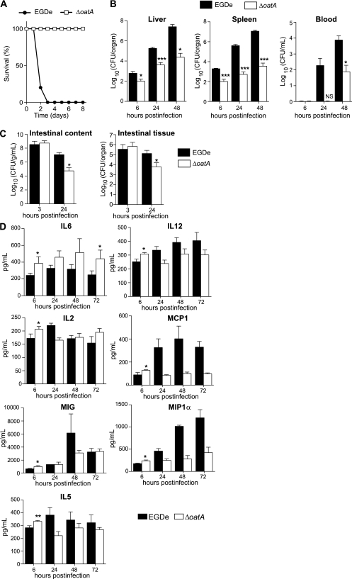
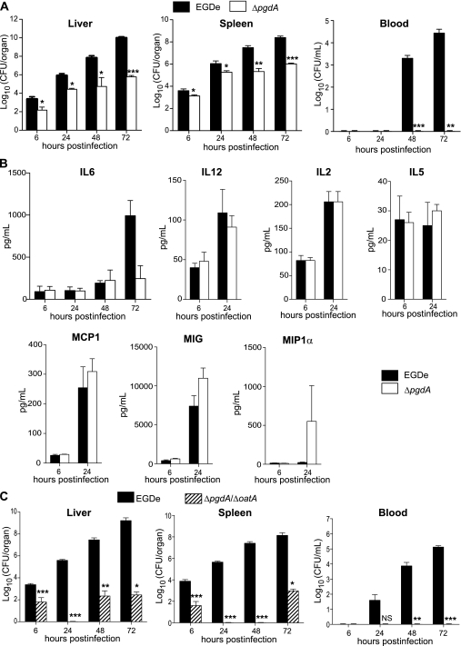
Microbial pathogens have evolved mechanisms to overcome immune responses and successfully infect their host. Here, we studied how Listeria monocytogenes evades immune detection by peptidoglycan (PGN) modification. By analyzing L. monocytogenes muropeptides, we detected O-acetylated muramic acid residues. We identified an O-acetyltransferase gene, oatA, in the L. monocytogenes genome sequence. Comparison of PGN from parental and isogenic oatA mutant strains showed that the O-acetyltransferase OatA O-acetylates Listeria PGN. We also found that PGN O-acetylation confers resistance to different types of antimicrobial compounds targeting bacterial cell wall such as lysozyme, β-lactam antibiotics, and bacteriocins and that O-acetylation is required for Listeria growth in macrophages. Moreover, oatA mutant virulence is drastically affected in mice following intravenous or oral inoculation. In addition, the oatA mutant induced early secretion of proinflammatory cytokines and chemokines in vivo. These results suggest an important role for OatA in limiting innate immune responses and promoting bacterial survival in the infected host.


Figures

Figure 1.
Figure 1.
Lmo1291 is a peptidoglycan O-acetyltransferase. A, Genetic organization of the L monocytogenes lmo1291 gene locus (in black) showing chromosomal coordinates (bp), putative terminators (black hairpins), and the flanking genes lmo1290 encoding a putative internalin (in white) and lmo1292 encoding a putative glycerophosphodiester phosphodiesterase (in gray). B, Map of putative domains of Lmo1291 (amino acid). C, Sequence alignment of O-acetyltransferases from Enterococcus faecalis (Efa), Lactococcus lactis (Lla), Staphylococcus aureus (Sau), and L. monocytogenes Lmo1291 (Lmo). Sequences were aligned using T-Coffee software. Amino acids in red are identical and residues in green are similar. Asterisks indicate the conserved catalytic triad of SGNH hydrolases. Efa, Lla, and Sau orthologs show 56%, 50%, and 55% identity to Lmo1291, respectively. D, Muropeptide profile of the highly purified cell walls of EGDe and its isogenic ΔoatA mutant. Peaks highlighted by an asterisk indicate muropeptides absent from the ΔoatA mutant. Major peaks were analyzed by MALDI MS/MS and their respective structure are indicated above the corresponding peak. Deacetylated muropeptides are indicated in red. Muropeptides that are O-acetylated are highlighted by the addition of the OAc moiety in blue. Dotted arrows indicate the relationship between the different O-acetylated muropeptides and their parent muropeptides that lack the OAc moiety. E, Localization of PGN modifications by OatA (O-acetylation in blue) and PgdA (N-deacetylation in red). MALDI indicates matrix-assisted laser desorption ionization; MS/MS, tandem mass spectrometry.
Figure 2.
Figure 2.
Inactivation of oatA increases sensitivity to antimicrobial molecules that target bacterial cell wall and impairs L monocytogenes survival in macrophages. A, Lysozyme disk-diffusion assay. Growth inhibition caused by lysozyme loaded on a paper disk (1 mg/disk) was measured on BHI agar plates inoculated with EGDe (black bars), the ΔoatA mutant (white bars), or ΔoatA+oatA−complemented strains (hatched bars). B, Cefotaxime minimum inhibitory concentration was determined using E-test strips on BHI agar plates that were inoculated with EGDe (black bars), ΔoatA mutant (white bars), or ΔoatA+oatA−complemented strains (hatched bars). C, Gallidermin inhibitory activity was determined in 96-well plates. 106 CFU/mL of EGDe (black bars) or ΔoatA (white bars) were incubated with increasing concentrations of gallidermin. The number of CFU in each well was assessed after overnight incubation at 37°C by plating serial dilutions on BHI agar plates. D, THP-1 cells (n = 4); E, peritoneal-elicited macrophages (n = 5); and F, bone marrow–derived macrophages (n = 4) were infected with EGDe (black circles) or ΔoatA (white squares). G, RAW264.7 cells (n = 5) were infected with EGDe (black circles), ΔoatA (white squares), or the ΔoatA+oatA complemented strain (gray triangles). The number of CFU was determined at different time points after cell lysis with 0.2% triton. Data are means ± SD. (HI) Electron microscopy analysis of RAW264.7 cells infected with EGDe or ΔoatA. (H) RAW264.7 cells after 4 hours of infection with EGDe or ΔoatA. Left panel: EGDe in vacuoles and protrusions; right panel: ΔoatA free in the cytosol. Scale bars: 2 μm. I, The number of bacteria per cell was determined by counting intravacuolar and cytosolic bacteria 30 minutes and 4 hours postinfection. Data are means ± SD (n = 25). Student t test was performed to determine statistical significance (*** indicates P < .001). CFU, colony-forming unit.
Figure 3.
Figure 3.
The ΔoatA mutant has a strongly attenuated virulence in mice and triggers an increased cytokine response early after infection. A, BALB/c mice were challenged by intravenous injection of 106 EGDe (black circles) or ΔoatA (white squares). Survival of infected mice was determined over time (n = 5). B, BALB/c mice were challenged by intravenous injection of a sublethal dose (104 bacteria per mouse) of EGDe (black bars) or ΔoatA (white bars). Colonization of liver, spleen, and blood was followed 6, 24, and 48 hours postinfection. Data are means ± SD (n = 4). C, Transgenic human E-cadherin mice were used as a permissive model for the oral route of infection. These mice were infected with 1010 EGDe (black bars) or ΔoatA (white bars). After 3 hours and 24 hours, mice were euthanized and the number of bacteria in the intestinal lumen and intestinal tissue was determined. Data are means ± SD (n = 3–5). D, BALB/c mice were inoculated intravenously with 5 × 10³ bacteria. The liver was dissected and homogenized. Homogenates were assayed using a multiplex immunoassay to determine cytokine level in response to infection with EGDe (black bars) or ΔoatA (white bars). Data are means ± SD (n = 4). Student t test was performed to determine statistical significance: *, **, and *** indicate P < .05, P < .01, and P < .001, respectively.
Figure 4.
Figure 4.
The ΔpgdA mutant, while highly attenuated in virulence, does not induce IL6 and is more virulent than a ΔpgdAΔoatA double mutant. A, BALB/c mice were challenged by intravenous injection of a sublethal dose (104 bacteria per mouse) of the parental EGDe strain (black bars) and its isogenic ΔpgdA mutant (white bars). Colonization of liver, spleen, and blood was followed 6, 24, 48, and 72 hours postinfection. Data are means ± SD (n = 4). B, BALB/c mice were inoculated intravenously with 5 × 10³ bacteria. The liver was dissected and homogenized. Homogenates were assayed to determine cytokine level in response to infection with the parental EGDe strain (black bars) or its isogenic ΔpgdA mutant (white bars). Data are means ± SD (n = 4). C, BALB/c mice were challenged by intravenous injection of a sublethal dose (104 bacteria per mouse) of the parental EGDe strain (black bars) and its isogenic ΔpgdAΔoatA mutant (hatched bars). Colonization of liver, spleen, and blood was followed at 6, 24, 48, and 72 hours postinfection. Data are means ± SD (n = 4). Student t test was performed to determine statistical significance: *, **, and *** indicate P < .05, P < .01, and P < .001, respectively.

References

    1. Vazquez-Boland JA, Kuhn M, Berche P, et al. Listeria pathogenesis and molecular virulence determinants. Clin Microbiol Rev. 2001;14:584–640. - PMC - PubMed
    1. Pizarro-Cerda J, Cossart P. Listeria monocytogenes membrane trafficking and lifestyle: the exception or the rule? Annu Rev Cell Dev Biol. 2009;25:649–70. - PubMed
    1. Hamon M, Bierne H, Cossart P. Listeria monocytogenes: a multifaceted model. Nat Rev Microbiol. 2006;4:423–34. - PubMed
    1. Pamer EG. Immune responses to Listeria monocytogenes. Nat Rev Immunol. 2004;4:812–23. - PubMed
    1. Stavru F, Archambaud C, Cossart P. Cell biology and immunology of Listeria monocytogenes infections: novel insights. Immunol Rev. 2011;240:160–84. - PubMed

Publication types

MeSH terms