. 2011 Aug 30;108(35):14643-8.
doi: 10.1073/pnas.1019074108. Epub 2011 Aug 15.

Conjugative DNA transfer into human cells by the VirB/VirD4 type IV secretion system of the bacterial pathogen Bartonella henselae

Affiliations

Conjugative DNA transfer into human cells by the VirB/VirD4 type IV secretion system of the bacterial pathogen Bartonella henselae

Gunnar Schröder et al. Proc Natl Acad Sci U S A. .

Abstract

Bacterial type IV secretion systems (T4SS) mediate interbacterial conjugative DNA transfer and transkingdom protein transfer into eukaryotic host cells in bacterial pathogenesis. The sole bacterium known to naturally transfer DNA into eukaryotic host cells via a T4SS is the plant pathogen Agrobacterium tumefaciens. Here we demonstrate T4SS-mediated DNA transfer from a human bacterial pathogen into human cells. We show that the zoonotic pathogen Bartonella henselae can transfer a cryptic plasmid occurring in the bartonellae into the human endothelial cell line EA.hy926 via its T4SS VirB/VirD4. DNA transfer into EA.hy926 cells was demonstrated by using a reporter derivative of this Bartonella-specific mobilizable plasmid generated by insertion of a eukaryotic egfp-expression cassette. Fusion of the C-terminal secretion signal of the endogenous VirB/VirD4 protein substrate BepD with the plasmid-encoded DNA-transport protein Mob resulted in a 100-fold increased DNA transfer rate. Expression of the delivered egfp gene in EA.hy926 cells required cell division, suggesting that nuclear envelope breakdown may facilitate passive entry of the transferred ssDNA into the nucleus as prerequisite for complementary strand synthesis and transcription of the egfp gene. Addition of an eukaryotic neomycin phosphotransferase expression cassette to the reporter plasmid facilitated selection of stable transgenic EA.hy926 cell lines that display chromosomal integration of the transferred plasmid DNA. Our data suggest that T4SS-dependent DNA transfer into host cells may occur naturally during human infection with Bartonella and that these chronically infecting pathogens have potential for the engineering of in vivo gene-delivery vectors with applications in DNA vaccination and therapeutic gene therapy.


Conflict of interest statement

The authors declare no conflict of interest.

Figures

Fig. 1.
Fig. 1.
B. henselae translocates plasmid DNA into human cells via the VirB/VirD4 T4SS. Ea.hy926 cells were infected with B. henselae WT or ΔvirB4 strains harboring the indicated plasmids. Plasmid transfer was monitored by measuring GFP fluorescence at 72 h of infection. (A) Schematic representation of the reporter plasmid pRS117 carrying an eukaryotic gfp-expression cassette (PCMV, egfp) plus plasmid mobilization factors (oriT, mob). Derivatives of this plasmid include a disruption of mob (pRGS06) and a fusion of the C-terminal BID domain of BepD to Mob (pRS122). (B) Percentage of gpc of infected cell populations, monitored by FACS analysis. The values indicated by an asterisk are below the detection limit (<0.003%). (C) Representative FACS profiles. FL-1, relative GFP fluorescence intensity; FSC, forward scatter. (D) Fluorescence microscopic picture (GFP channel) of a cell preparation infected with B. henselae WT carrying pRS122. The image was overlaid with a phase contrast picture of the same microscopic field. (Scale bar: 10 μm.) (E) Model view of the B. henselae VirB/VirD4 T4SS apparatus transporting plasmid DNA into a human host cell. Eleven components, including the ATPases VirB4, VirB11, and VirD4 (indicated as B4, B11, and D4, respectively), form a channel-spanning inner membrane (IM), periplasm (PP), peptidoglycan (PG), and outer membrane (OM) of the bacterium, plus the cellular membrane (CM) of the human host cell.
Fig. 2.
Fig. 2.
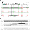
Integration of translocated plasmid DNA into the host cell chromosome. Following infection of Ea.hy926 cells with B. henselae strain RSE581 carrying the reporter plasmid pRS130, eight individual cell lines (named A–H) where isolated. (A) Schematic representation shows which parts of plasmid pRS130 have integrated into the genome of recombinant cell lines A–H, based on Southern blot analysis (Fig. S1 and SI Results) and PCR analysis (Table S1). A total of 13 different primer pairs (numbered 1–13) were tested on each cell line for presence (green box) or absence (red open box) of the targeted sequence. Note that the leftmost amplicon spans the oriT of pRS130. The border presented in B is indicated by an asterisk and the borders presented in Fig. S2 are marked by a number sign (#). (B) Nucleotide sequence alignment of the oriT region of pRS130, the isolated integration junction of the transferred DNA [line C (5′)], and the human DNA sequence (accession no. AC108035). The oriT of pRS130 [as defined in the parental plasmid pBGR1 (20)] is highlighted in black, and the start codon of the mob gene is marked by three asterisks. Homology of the junction fragment to pRS130 or AC108035 is indicated by open boxes. In the 47-bp spacer region that does not show colinearity to either donor or recipient sequence, the 22 nucleotides upstream from the oriT are found as an inverted element (gray arrow). Homologies to pRS130 or AC108035 within these 47 bp are indicated; 240 bp of the isolated fragment, centered around the integration site, are displayed.
Fig. 3.
Fig. 3.
Expression of a delivered egfp gene requires cell division. GFP expression resulting from DNA transfer or protein transfer was measured as a function of initial cell density or as a function of time post infection as percent gpc. (A) Host cells were seeded into 24-well plates at increasing cell densities (indicated in cells per well) to obtain different degrees of confluence. Bacterial strains RSE556 (DNA transfer) or RSE308 (protein transfer by Cre recombinase assay for translocation) were added at a multiplicity of infection of 200 and percent gpc levels were measured after 3 d. (B) Host cells (80,000 cells per well) were infected as before and percent gpc levels were monitored between day 1 and day 6 after infection.

References

    1. Alvarez-Martinez CE, Christie PJ. Biological diversity of prokaryotic type IV secretion systems. Microbiol Mol Biol Rev. 2009;73:775–808. - PMC - PubMed
    1. Frank AC, Alsmark CM, Thollesson M, Andersson SG. Functional divergence and horizontal transfer of type IV secretion systems. Mol Biol Evol. 2005;22:1325–1336. - PubMed
    1. Schröder G, Lanka E. The mating pair formation system of conjugative plasmids-A versatile secretion machinery for transfer of proteins and DNA. Plasmid. 2005;54:1–25. - PubMed
    1. Bates S, Cashmore AM, Wilkins BM. IncP plasmids are unusually effective in mediating conjugation of Escherichia coli and Saccharomyces cerevisiae: involvement of the tra2 mating system. J Bacteriol. 1998;180:6538–6543. - PMC - PubMed
    1. Heinemann JA, Sprague GF., Jr Bacterial conjugative plasmids mobilize DNA transfer between bacteria and yeast. Nature. 1989;340:205–209. - PubMed

Publication types

Substances