. 2011 Dec 20;108(51):20325-32.
doi: 10.1073/pnas.1100279108. Epub 2011 Aug 15.

Architecture of human telomerase RNA

Affiliations

Architecture of human telomerase RNA

Qi Zhang et al. Proc Natl Acad Sci U S A. .

Abstract

Telomerase is a unique reverse transcriptase that catalyzes the addition of telomere DNA repeats onto the 3' ends of linear chromosomes and plays a critical role in maintaining genome stability. Unlike other reverse transcriptases, telomerase is unique in that it is a ribonucleoprotein complex, where the RNA component [telomerase RNA (TR)] not only provides the template for the synthesis of telomere DNA repeats but also plays essential roles in catalysis, accumulation, TR 3'-end processing, localization, and holoenzyme assembly. Biochemical studies have identified TR elements essential for catalysis that share remarkably conserved secondary structures across different species as well as species-specific domains for other functions, paving the way for high-resolution structure determination of TRs. Over the past decade, structures of key elements from the core, conserved regions 4 and 5, and small Cajal body specific RNA domains of human TR have emerged, providing significant insights into the roles of these RNA elements in telomerase function. Structures of all helical elements of the core domain have been recently reported, providing the basis for a high-resolution model of the complete core domain. We review this progress to determine the overall architecture of human telomerase RNA.


Conflict of interest statement

The authors declare no conflict of interest.

Figures
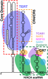
Fig. 1.
Fig. 1.
Secondary structure and known protein components of the human telomerase holoenzyme. The human telomerase RNA (hTR) contains three major structural and functional domains, the core domain, the CR4/CR5 domain, and the H/ACA scaRNA domain (46, 59, 62). The hTR core and CR4/CR5 domains independently bind the hTERT (blue ellipse) (50, 64). The hTR scaRNA domain binds two sets of the four H/ACA RNP proteins: dyskerin (green), Gar1 (cyan), Nop10 (magenta), and Nhp2 (orange) (67). The protein TCAB1/WDR79 (purple) binds both the dyskerin and the CAB box located at the CR7 region within the H/ACA scaRNA domain (72, 73).
Fig. 2.
Fig. 2.
Structures of subdomains of the hTR core domain. (A) Sequence and secondary structure of the hTR core domain, which can be divided into four subdomains: P2a-J2a.1-P2a.1 (blue-gray-gold), P2a-J2a/b-P2b (gold-green-red), P2b-P3 pseudoknot (red-pink), and P1 (dark green) (46). (B) NMR solution structures of the P2b-P3 pseudoknot, where PKDU (PDB ID 2K96) and PKWT (PDB ID 2K96) are structures for ΔU177 mutant and WT constructs, respectively (54). (C) NMR solution structure of P2a-J2a/b-P2b (PDB ID 2L3E) (55). (D) Structural model of the P2a-J2a.1-P2a.1 determined by the RDC-MC-Sym approach (55). All 3D structures are color-coded like the secondary structure in A, and nonnative residues are colored light gray.
Fig. 3.
Fig. 3.
Tertiary interactions in the pseudoknot. (A) The structure of the triple helical region of PKWT (54). (B) Schematic representations of the tertiary interactions in PKWT. In PKDU, U103 forms a Hoogsteen base pair with G178. (C) U·A-U Hoogsteen base triple. (D) Detailed view of the base pairs surrounding U177. The base of U177 stacks over the 2′OH of A176. The nucleotides are in CPK colors, except that U177 is colored magenta.
Fig. 4.
Fig. 4.
Models of the hTR core domain and interaction with TERT. (A) NMR-based model of the hTR core domain including a DNA primer bound to the template (55). (B) The hTR P2/P3 pseudoknot positioned onto the T. castaneum TERT in two possible orientations, where the hTR P2/P3 pseudoknot lies either parallel (Left) or perpendicular (Right) to the T. castaneum TERT-telomeric RNA/DNA complex (PDB ID 3KYL) (37). The color scheme for the hTR P2/P3 pseudoknot domain is the same as in Fig. 2. Domains of the T. castaneum TERT are colored and labeled as shown, and the RNA template and telomeric DNA are colored in purple and cyan, respectively. A comparison of the domain structures of the hTERT and T. castaneum TERT is also shown.
Fig. 5.
Fig. 5.
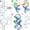
Structures of subdomains of the hTR CR4/CR5 domain. (A) Sequence and secondary structure of the hTR CR4/CR5 domain (46). The minimal CR4/CR5 required for reconstitution in vitro of active telomerase is highlighted by a dashed box (76). (B) NMR solution structures of P6.1 (PDB ID 1OQ0) (75) and Ψ4P6.1 (PDB ID 2KYE) (76). The two U and three G letters in the P6.1 loop are colored in green and yellow, respectively. The two pseudouridines (Ψs) in the P6.1 loop are colored in red. (C) NMR solution structure and secondary structure of a P6 hairpin construct (PDB ID 1Z31) (74). The internal loop residues (C266-C267 and A289-U291) are colored in green, and the bulge residue C262 is colored in purple.
Fig. 6.
Fig. 6.
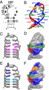
Structure and function of the hTR CR7 domain (60). (A) Sequence and secondary structure of the hTR CR7 hairpin and flanking base pairs. Boxed residues are the CAB box sequence. Residues with >95% and >85–95% conservation among all vertebrate species are shown in capital letters and bold fonts, respectively. Nonnative residues used in structure determination are the base pairs below the dashed line. (B) NMR solution structure of the CR7 hairpin (PDB ID 2QH2). Residues are colored by type: orange, A; green, U; blue, G; red, C. (C and D) Residues comprising the hTR-specific processing signal are highlighted in magenta in the secondary structure (C) and colored as in B in the surface representation of the CR7 structure (D). (E and F) Residues comprising the CB localization signal are highlighted in dark blue (CAB box) and light blue in the secondary structure (E) and colored as in B in the surface representation of the CR7 structure (F).

References

    1. Palm W, de Lange T. How shelterin protects mammalian telomeres. Annu Rev Genet. 2008;42:301–334. - PubMed
    1. Martínez P, Blasco MA. Telomeric and extra-telomeric roles for telomerase and the telomere-binding proteins. Nat Rev Cancer. 2011;11:161–176. - PubMed
    1. Wong JM, Collins K. Telomere maintenance and disease. Lancet. 2003;362:983–988. - PubMed
    1. Blasco MA. Telomere length, stem cells and aging. Nat Chem Biol. 2007;3:640–649. - PubMed
    1. Collado M, Blasco MA, Serrano M. Cellular senescence in cancer and aging. Cell. 2007;130:223–233. - PubMed

Publication types

LinkOut - more resources