. 2011 Aug 30;108(35):14473-8.
doi: 10.1073/pnas.1110412108. Epub 2011 Aug 15.

Augmin promotes meiotic spindle formation and bipolarity in Xenopus egg extracts

Affiliations

Augmin promotes meiotic spindle formation and bipolarity in Xenopus egg extracts

Sabine Petry et al. Proc Natl Acad Sci U S A. .

Abstract

Female meiotic spindles in many organisms form in the absence of centrosomes, the organelle typically associated with microtubule (MT) nucleation. Previous studies have proposed that these meiotic spindles arise from RanGTP-mediated MT nucleation in the vicinity of chromatin; however, whether this process is sufficient for spindle formation is unknown. Here, we investigated whether a recently proposed spindle-based MT nucleation pathway that involves augmin, an 8-subunit protein complex, also contributes to spindle morphogenesis. We used an assay system in which hundreds of meiotic spindles can be observed forming around chromatin-coated beads after introduction of Xenopus egg extracts. Spindles forming in augmin-depleted extracts showed reduced rates of MT formation and were predominantly multipolar, revealing a function of augmin in stabilizing the bipolar shape of the acentrosomal meiotic spindle. Our studies also have uncovered an apparent augmin-independent MT nucleation process from acentrosomal poles, which becomes increasingly active over time and appears to partially rescue the spindle defects that arise from augmin depletion. Our studies reveal that spatially and temporally distinct MT generation pathways from chromatin, spindle MTs, and acentrosomal poles all contribute to robust bipolar spindle formation in meiotic extracts.


Conflict of interest statement

The authors declare no conflict of interest.

Figures
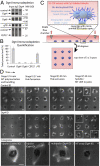
Fig. 1.
Fig. 1.
Characterization of acentrosomal spindles from Dgt4- and control-depleted extracts around microprinted DNA beads in a regular pattern. (A) Immunoblot of immunodepletion of Dgt4 and total IgG fraction antibodies as a control. The augmin subunits Dgt4, Dgt6, and CEP27 were specifically codepleted from the extract. (B) Quantification of Dgt4 immunodepletions from at least three independent experiments. The mean percentage is relative to the input material ± SD. (C) Assay for spindle formation around chromatin-coated beads. Biotinylated DNA beads were attached to microprinted BSA–biotin spots via streptavidin magnetic beads (17). (D) Images of a 3 × 3 DNA bead pattern during spindle assembly for the control-depleted (Top) and Dgt4-depleted (Middle) extracts. The characteristic stages of spindle formation are depicted. The 3 × 3 DNA bead pattern of the Dgt4-depleted extract was obtained from an individual field of view. In contrast, the pattern of the control-depleted extract is from adjacent fields of view, and therefore contains border lines. (Bottom) Examples of the three spindle categories (bipolar, multipolar, and MT array; from the boxes in the 3 × 3 arrays). (Scale bars, 10 μm.) Effects of γ-tubulin depletion are shown in Fig. S2.
Fig. 2.
Fig. 2.
Kinetics of spindle assembly in control- and Dgt4-depleted extracts. The background-subtracted fluorescent tubulin intensity around individual DNA bead spots was measured at regular time intervals (for each time point, ≤81 chromatin beads were averaged and normalized to the maximum TB intensity of the individual experiment). Results from three independent experiments with different extracts are shown (circles, squares, and crosses). The combined data were fit to a model with two ordinary differential equations using four parameters (see ref. 17; Fig. 4) and is depicted as a dotted line. The relative total tubulin intensity between control- and augmin-depleted structures was scaled based on a representative experiment. The onset of MT nucleation varied slightly with each extract, but occurred on average 11 min later for the Dgt4-depleted extract. The timing of stages I–IV shown in Fig 1B are indicated on the graph.
Fig. 3.
Fig. 3.
Characteristics of acentrosomal spindles from Dgt4- and control-depleted spindles. (A) Quantification of control-depleted (Upper) and Dgt4-depleted (Lower) acentrosomal structures (mean and SEM shown; three independent experiments with n ≥ 100 measurements each at each time point). See Fig. 1 for classification of bipolar, multipolar, and MT array. Quantitation of spindle length (B), total MT intensity (C), and the ratio between MT intensity in the midzone and near the poles (D) are shown. Mean and SEM are shown from mean results of three independent experiments (n > 15 for each experiment). (E) Alignment of poles in bipolar spindles from control and augmin-depleted extracts. The angle between one pole, the middle of the DNA bead spot, and the second pole were measured. An angle of 180° reflects a perfectly symmetric spindle. (F) Quantification of the pole–DNA–pole angle for structures of control- and Dgt4-depleted extracts (mean and SD from three independent experiments; n = 20 measurements from each experiment). The pole–DNA–pole angle in Dgt4-depleted extracts was significantly (P < 0.001) lower compared with control-depleted extracts. (G) Images of a 3 × 3 DNA bead pattern during acentrosomal spindle assembly and quantification of acentrosomal structures for extracts supplemented with control antibody (Left) or Dgt4 antibody (Right) (77.5 μg/mL antibody added at time 0). The 3 × 3 DNA bead pattern with the Dgt4 antibody was obtained from an individual field of view. In contrast, the pattern with the control antibody is from adjacent fields of view, and therefore contains border lines. Data at each time point were derived from three independent experiments (mean and SEM are shown). (Scale bars, 10 μm.)
Fig. 4.
Fig. 4.
Visualization of growing MT plus ends via GFP-EB1 in extracts with control antibody (Left) or anti-Dgt4 antibody (Right). (A) Representative images of tubulin (Cy3-channel) and GFP–EB1 (140 nM) are shown for extract incubated with control or Dgt4 antibody (see Fig. 3G and Fig. S3 CE) at three assembly stages (see Fig. 1D). (B and C) Augmin-inhibited spindles show a reduction of EB1–GFP comets from the middle of the spindles. Note the prominent density of EB1–GFP comets at the acentrosomal poles in C (both control and Dgt4 AB). (Scale bars, 10 μm.) See also Movie S3 (control AB) and Movie S4 (Dgt4 AB). (D) Quantitation of the ratio between the EB1 intensity in the midzone and near the poles (see circles for zone of measurement). Mean and SEM are derived from three independent experiments (2–7 spindles measured for each time point in each independent experiment).
Fig. 5.
Fig. 5.
Models of MT nucleation and spindle assembly with and without augmin. (A) MT nucleation occurs first around chromatin and is then amplified by augmin, leading to straight and rigid MT bundles. Many of the bundles extend MTs that cross the DNA midzone, contributing to a zone of antiparallel MTs. (B) The Kinesin-5 motor Eg5 pushes antiparallel MTs apart, whereas dynein moves toward and gathers MT minus ends together, thus contributing to pole formation. (C) A bipolar spindle is formed with two foci of MT minus ends and antiparallel MT overlap in the central body of the spindle. MT nucleation from the two acentrosomal poles reinforces the spindle bipolarity. (D) Without augmin, MTs are generated near chromatin, but subsequent MT amplification and the formation of a functional overlap zone are impaired. (E) Dynein gathers MT minus ends into multiple poles, due to the lack of an overlap zone and possibly due to loss of augmin-mediated MT crosslinking. (F) At late time points, nucleation activity shifts to the poles, similar to wild-type spindles. This shift facilitates production of MTs and may help to rescue spindle bipolarity in some cases. However, the lack of a strong antiparallel MT overlap in the middle leads to a weakly connected and elongated spindle structure.

References

    1. Wiese C, Zheng YX. Microtubule nucleation: gamma-tubulin and beyond. J Cell Sci. 2006;119:4143–4153. - PubMed
    1. Kollman JM, Polka JK, Zelter A, Davis TN, Agard DA. Microtubule nucleating gamma-TuSC assembles structures with 13-fold microtubule-like symmetry. Nature. 2010;466:879–882. - PMC - PubMed
    1. Mitchison T, Kirschner M. Dynamic instability of microtubule growth. Nature. 1984;312:237–242. - PubMed
    1. Khodjakov A, Cole RW, Oakley BR, Rieder CL. Centrosome-independent mitotic spindle formation in vertebrates. Curr Biol. 2000;10:59–67. - PubMed
    1. Megraw TL, Kao LR, Kaufman TC. Zygotic development without functional mitotic centrosomes. Curr Biol. 2001;11:116–120. - PubMed

Publication types