. 2011 Aug 30;108(35):14497-502.
doi: 10.1073/pnas.1111309108. Epub 2011 Aug 15.

OCT4 establishes and maintains nucleosome-depleted regions that provide additional layers of epigenetic regulation of its target genes

Affiliations

OCT4 establishes and maintains nucleosome-depleted regions that provide additional layers of epigenetic regulation of its target genes

Jueng Soo You et al. Proc Natl Acad Sci U S A. .

Abstract

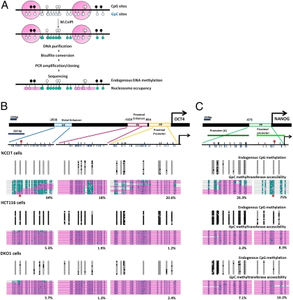
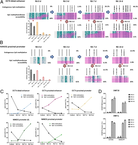
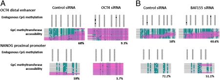
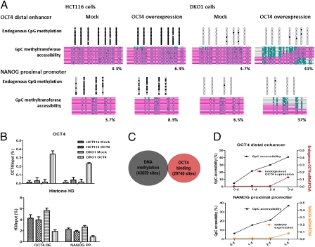
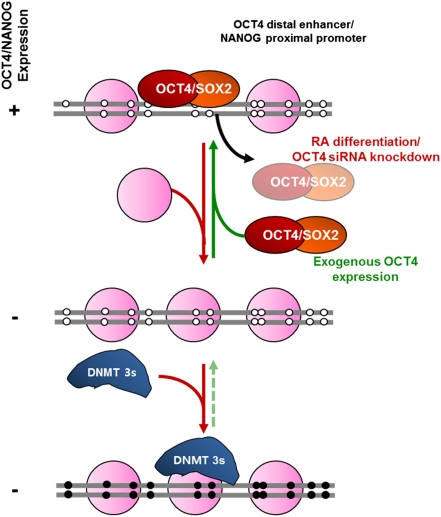
Recent epigenome-wide mapping studies describe nucleosome-depleted regions (NDRs) at transcription start sites and enhancers. However, these static maps do not address causality or the roles of NDRs in gene control, and their relationship to transcription factors and DNA methylation is not well understood. Using a high-resolution single-molecule mapping approach to simultaneously investigate endogenous DNA methylation and nucleosome occupancies on individual DNA molecules, we show that the unmethylated OCT4 distal enhancer has an NDR, whereas NANOG has a clear NDR at its proximal promoter. These NDRs are maintained by binding of OCT4 and are required for OCT4 and NANOG expression. Differentiation causes a rapid loss of both NDRs accompanied by nucleosome occupancy, which precedes de novo DNA methylation. NDRs can be restored by forced expression of OCT4 in somatic cells but only when there is no cytosine methylation. These data show the central role of the NDRs, established by OCT4, in ensuring the autoregulatory loop of pluripotency and, furthermore, that de novo methylation follows the loss of NDRs and stabilizes the suppressed state.


Conflict of interest statement

The authors declare no conflict of interest.

Figures

Fig. 1.
Fig. 1.
OCT4 and NANOG show distinct nucleosome configurations at DNA regulatory regions that correlate with transcriptional activities. (A) Scheme of the NOMe-seq assay. Upper black circles represent CpG sites (white circles represent unmethylated CpG sites, and black circles represent methylated CpG sites). Lower blue circles represent GpC sites (unfilled blue circles represent GpC sites that are inaccessible to GpC methyltransferase, and green-filled blue circles represent sites that are accessible to GpC methyltransferase). Pink bars represent regions of inaccessibility large enough to accommodate a nucleosome. (B and C) Schematic diagrams of the human OCT4 distal enhancer (DE), proximal enhancer (PE), and proximal promoter (PP) and the NANOG promoter. OCT4 recognizes specific octamer sequences at the OCT4 distal enhancer and NANOG proximal promoter (denoted by asterisks). Arrows indicate the transcription start sites. Small upper black lines indicate CpG sites, and lower blue lines indicate GpC sites of those regions. Endogenous DNA methylation and nucleosome occupancy were analyzed by the NOMe-seq assay. A 150-bp marker gives the approximate size that would accommodate a nucleosome. Each horizontal line represents individual OCT4 enhancers and promoter or NANOG promoter modules.
Fig. 2.
Fig. 2.
DNA methylation follows nucleosome insertion during cell differentiation and inhibits reactivation of OCT4 and NANOG gene expression. NCCIT cells were exposed to 10 μM RA for the indicated days. (A and B) Endogenous DNA methylation and nucleosome occupancy at the OCT4 distal enhancer and NANOG proximal promoter were analyzed by the NOMe-seq assay during differentiation. OCT4 recognizes specific octamer sequences at the OCT4 distal enhancer and NANOG proximal promoter that do not include CpG sites (denoted by asterisks). For reactivation experiments, RA was removed and cells were cultured for an additional 3 d with fresh growth media. The expression levels of OCT4 and NANOG were determined by quantitative PCR at each indicated time point. (C) Kinetics of the change in DNA methylation and nucleosome occupancy at the OCT4 and NANOG DNA regulatory regions. (D) Chromatin was immunoprecipitated with anti-DNMT3B and anti-DNMT3L antibodies at each time point during the differentiation, and their binding to the OCT4 distal enhancer and NANOG proximal promoter was analyzed by quantitative PCR.
Fig. 3.
Fig. 3.
OCT4 maintains NDRs at regulatory regions of OCT4 and NANOG, which ensures the autoregulatory loop of pluripotency. (A) At 72 h posttransfection with OCT4 and control siRNAs (100 nmol/L), endogenous DNA methylation and nucleosome occupancy at the OCT4 distal enhancer and NANOG proximal promoter were analyzed by the NOMe-seq assay. The data are representative of three biological experiments. (B) At 72 h posttransfection with BAF155 or OCT4 siRNA in NCCIT cells, the NOMe-seq assay was performed. The data are representative of two biological experiments.
Fig. 4.
Fig. 4.
Exogenous OCT4 expression initiates nucleosome depletion at DNA regulatory regions of OCT4 and NANOG, which are inhibited by DNA methylation. (A) Transfection of HCT116 and DKO1 cells with exogenous OCT4 and mock vectors was carried out with Lipofectamine LTX. At 72 h posttransfection, endogenous DNA methylation and nucleosome occupancy at the OCT4 distal enhancer and NANOG proximal promoter were analyzed by the NOMe-seq assay. (B) At the same time points, ChIP assays were performed with OCT4 and histone H3. (C) To understand the relationship between OCT4 binding and DNA methylated regions, genome-wide studies in human embryonic stem cells (H1) using ENCODE and GEO data (wgEncodeHudsonalphaMethylSeqRegionsRep1H1hesc for DNA methylation and GSM518373 for OCT4 ChIP-Seq) were performed. The data comprised 100-bp windows of OCT4 binding regions (29,740 sites) and DNA methylation (43,659 sites). (D) DKO1 cells were transfected with exogenous OCT4 and mock vectors. The NOMe-seq assay was performed at the indicated time after transfection.
Fig. 5.
Fig. 5.
A proposed model of the mechanism of repression and de novo DNA methylation of OCT4 and NANOG during cell differentiation. (Refer to Discussion for a detailed explanation.)

References

    1. Reik W. Stability and flexibility of epigenetic gene regulation in mammalian development. Nature. 2007;447:425–432. - PubMed
    1. Hochedlinger K, Plath K. Epigenetic reprogramming and induced pluripotency. Development. 2009;136:509–523. - PMC - PubMed
    1. Sharma S, Kelly TK, Jones PA. Epigenetics in cancer. Carcinogenesis. 2010;31:27–36. - PMC - PubMed
    1. Christophersen NS, Helin K. Epigenetic control of embryonic stem cell fate. J Exp Med. 2010;207:2287–2295. - PMC - PubMed
    1. Bernstein BE, et al. A bivalent chromatin structure marks key developmental genes in embryonic stem cells. Cell. 2006;125:315–326. - PubMed

Publication types