. 2011 Aug 30;108(35):14614-9.
doi: 10.1073/pnas.1012859108. Epub 2011 Aug 15.

Vascular cell-adhesion molecule-1 plays a central role in the proangiogenic effects of oxidative stress

Affiliations

Vascular cell-adhesion molecule-1 plays a central role in the proangiogenic effects of oxidative stress

Aling Dong et al. Proc Natl Acad Sci U S A. .

Abstract

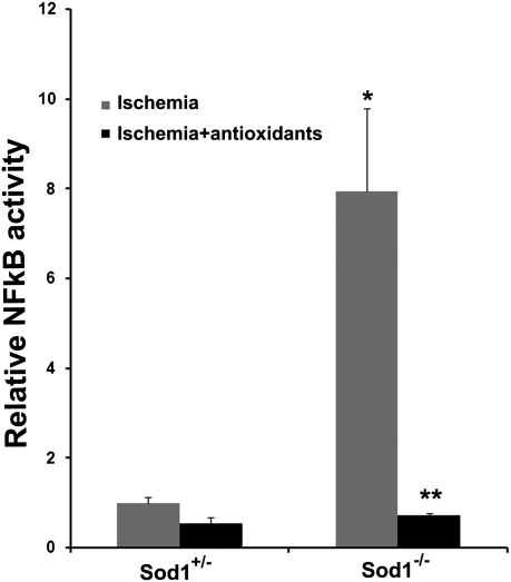
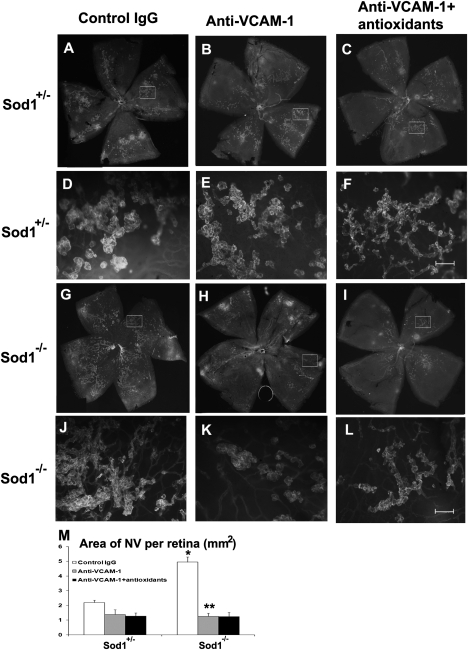
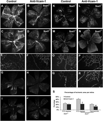
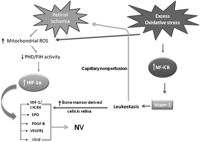
Oxidative stress exacerbates neovascularization (NV) in many disease processes. In this study we investigated the mechanism of that effect. Mice deficient in superoxide dismutase 1 (Sod1(-/-) mice) have increased oxidative stress and show severe ocular NV that is reduced to baseline by antioxidants. Compared with wild-type mice with ischemic retinopathy, Sod1(-/-) mice with ischemic retinopathy had increased expression of several NF-κB-responsive genes, but expression of vascular cell-adhesion molecule-1 (Vcam1) was particularly high. Intraocular injection of anti-VCAM-1 antibody eliminated the excessive ischemia-induced retinal NV. Elements that contributed to oxidative stress-induced worsening of retinal NV that were abrogated by blockade of VCAM-1 included increases in leukostasis, influx of bone marrow-derived cells, and capillary closure. Compared with ischemia alone, ischemia plus oxidative stress resulted in increased expression of several HIF-1-responsive genes caused in part by VCAM-1-induced worsening of nonperfusion and, hence, ischemia, because anti-VCAM-1 significantly reduced the increased expression of all but one of the genes. These data explain why oxidative stress worsens ischemia-induced retinal NV and may be relevant to other neovascular diseases in which oxidative stress has been implicated. The data also suggest that antagonism of VCAM-1 provides a potential therapy to combat worsening of neovascular diseases by oxidative stress.


Conflict of interest statement

The authors declare no conflict of interest.

Figures

Fig. 1.
Fig. 1.
Oxidative stress combined with ischemia up-regulates NF-κB activity in the retina. Sod1−/− and Sod1+/− pups were placed in 75% oxygen at P7, returned to room air at P12, and treated with antioxidants or vehicle (n = 5 for each group). At P15, NF-κB activity was measured in 10 μg of nuclear protein from each retina. There was a significant increase in NF-κB activity in the ischemic retinas of Sod1−/− vs. Sod1+/− mice (*P < 0.0001 by ANOVA with Dunnett's correction for multiple comparisons) that was completely blocked by antioxidants (**P < 0.0001).
Fig. 2.
Fig. 2.
Blockade of VCAM-1 eliminates the oxidative stress-induced increase of retinal NV in ischemic retina. Sod1−/− and Sod1+/− pups with ischemic retinopathy had intraocular injection of 1 μg of rat antimurine VCAM-1 in one eye and 1 μg of rat IgG in the fellow eye. At P17, in vivo immunostaining for PECAM-1, which selectively stains NV on the surface of the retina and hyaloid vessels; the hyaloid vessels are easily distinguished from NV because they are large diameter vessels. Eyes from Sod1+/− mice showed moderate NV on the surface of the retina that was similar in eyes that had been injected with IgG (A and D) or anti–VCAM-1 without (B and E) or with antioxidant treatment (C and F). Eyes from Sod1−/− mice that had been injected with IgG showed extensive NV on the surface of the retina (G and J), but Sod1−/− mice that had been injected with anti–VCAM-1 showed little NV (H and K) that was not further reduced by antioxidants (I and L). The mean (± SEM) area of retinal NV measured by image analysis (M) (n = 6 for each group) was significantly greater in IgG-injected eyes of Sod1−/− vs. Sod1+/− mice with ischemic retinopathy (*P < 0.0001 by ANOVA with Dunnett's correction for multiple comparisons), and also significantly greater (**P < 0.0001) than in anti–VCAM-1–injected eyes of Sod1−/− mice with ischemic retinopathy. [Magnification: A–C and G–I, 25× (images were taken at 25× and assembled into single image with Photoshop; DF and JL, 200×; Scale bar, 100 μm.]
Fig. 3.
Fig. 3.
The percentage area of nonperfused/hypoxic retina is increased in Sod1−/− mice versus Sod1+/− with ischemic retinopathy and the increase is blocked by anti–VCAM-1 antibody. Sod1−/− and Sod1+/− mice with ischemic retinopathy received an intraocular injection of 1 μg of anti–VCAM-1 in one eye and 1 μg of control IgG or BSA in the fellow eye. At P15, perfusion with FITC-labeled dextran showed that retinas from Sod1+/− mice injected with BSA (A) or anti–VCAM-1 (B) showed similar areas of nonperfusion, but retinas from Sod1−/− injected with BSA (C) or IgG (K) showed much larger areas of nonperfusion than those from eyes injected with anti–VCAM-1 (D and L). (E and F) High magnification of the boxed area at the border of perfused and nonperfused retina in C and D stained with GSA lectin labeled with Alexa594. There are nonperfused vessels (arrows) in the retina of BSA-injected eyes (E) not seen in anti–VCAM-1–injected eyes (F). The retinas of Sod1−/− injected with IgG showed larger areas of staining with hypoxyprobe (G), corresponding to larger areas of nonperfusion (I) compared with those injected with anti–VCAM-1 (H and J). FITC-dextran perfused retinas (K and L) stained for collagen IV (M and N) from P15 Sod1−/− mice with ischemic retinopathy showed larger avascular areas in IgG-injected eyes (K and M) than anti–VCAM-1–injected eyes (L and N). High magnification of boxed areas in M and N show nonperfused vascular stuctures in IgG injected eyes (Q vs. O, arrows) but not anti–VCAM-1–injected eyes (R vs. P). Image analysis showed that the mean (± SEM) percentage area of nonperfusion was greater in Sod1−/− mice compared with Sod1+/− mice and was significantly reduced by intraocular injection of anti–VCAM-1 (S) (*P = 0.0044 by ANOVA with Dunnett's correction for multiple comparisons). Furthermore, the mean area of hydroxyprobe-stained retina was significantly greater in Sod1−/− mice injected with control IgG than those injected with anti–VCAM-1 (S, Right) (**P = 0.0119). [Magnification: A–D, G, H, I, J, and K–N, 25× (images were taken at 25× and assembled into a single image with Photoshop); E, F, O, P, Q, and R, 200×; Scale bar, 100 μm.]
Fig. 4.
Fig. 4.
Mechanisms by which oxidative stress enhances NV in ischemic retina. Ischemia leads to stabilization of HIF-1 at least in part by generation of ROS that reduce activity of PHD. The stabilized HIF-1 translocates to the nucleus and increases expression of hypoxia-inducible genes, which cause neovascularization. If oxidative stress is added, there is activation of NF-κB, which increases expression several genes but results in particularly high levels of VCAM-1, which promotes leukostasis and influx of bone marrow-derived cells. Leukostasis causes leukocytic plugging, increases capillary nonperfusion, and worsens retinal ischemia, which elevates HIF-1 levels. The further elevation of HIF-1 enhances the already increased expression of HIF-1–responsive genes, which stimulates angiogenesis. Oxidative stress may also increase HIF-1 levels directly by ROS-mediated suppression of PHD. The increase in bone marrow-derived cells in the retina also release VEGF and other factors that further stimulate neovascularization.

Similar articles

Cited by

References

    1. Semenza GL. HIF-1: Mediator of physiological and pathophysiological responses to hypoxia. J Appl Physiol. 2000;88:1474–1480. - PubMed
    1. Ozaki H, et al. Hypoxia inducible factor-1α is increased in ischemic retina: Temporal and spatial correlation with VEGF expression. Invest Ophthalmol Vis Sci. 1999;40:182–189. - PubMed
    1. Kelly BD, et al. Cell type-specific regulation of angiogenic growth factor gene expression and induction of angiogenesis in nonischemic tissue by a constitutively active form of hypoxia-inducible factor 1. Circ Res. 2003;93:1074–1081. - PubMed
    1. Vinores SA, et al. Implication of the hypoxia response element of the Vegf promoter in mouse models of retinal and choroidal neovascularization, but not retinal vascular development. J Cell Physiol. 2006;206:749–758. - PubMed
    1. Aiello LP, et al. Suppression of retinal neovascularization in vivo by inhibition of vascular endothelial growth factor (VEGF) using soluble VEGF-receptor chimeric proteins. Proc Natl Acad Sci USA. 1995;92:10457–10461. - PMC - PubMed

Publication types

LinkOut - more resources