. 2011 Sep 15;187(6):3026-32.
doi: 10.4049/jimmunol.1101262. Epub 2011 Aug 15.

EBI2 guides serial movements of activated B cells and ligand activity is detectable in lymphoid and nonlymphoid tissues

Affiliations

EBI2 guides serial movements of activated B cells and ligand activity is detectable in lymphoid and nonlymphoid tissues

Lisa M Kelly et al. J Immunol. .

Abstract

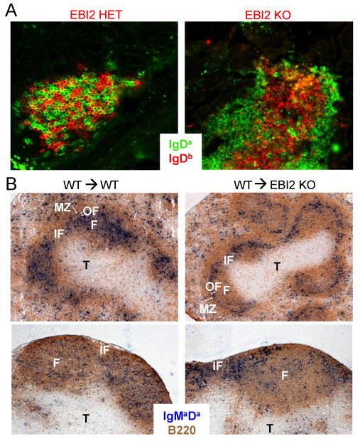
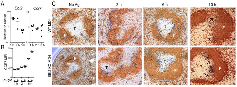
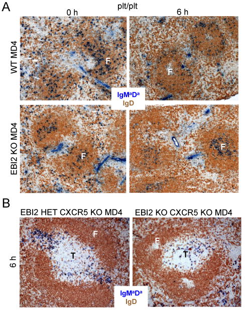
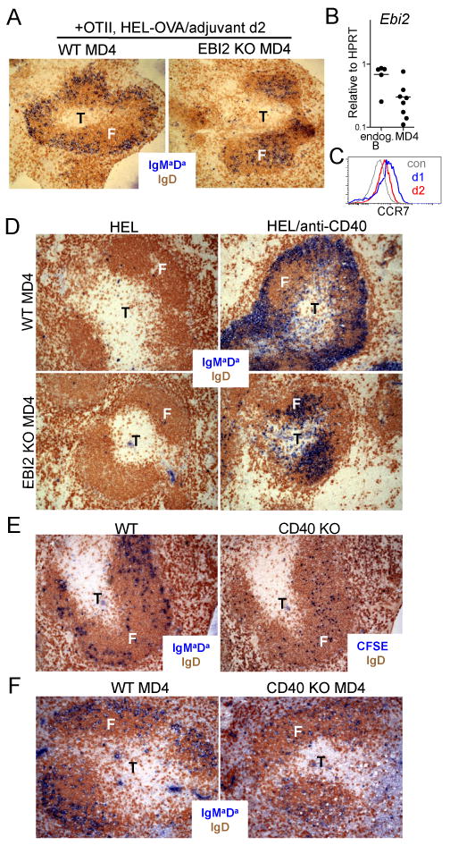
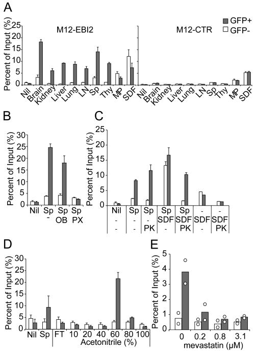
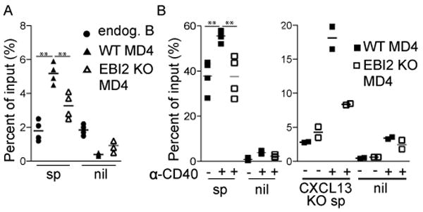
EBV-induced gene 2 (EBI2) was recently shown to direct the delayed movement of activated B cells to interfollicular and outer follicular regions of secondary lymphoid organs and to be required for mounting a normal T-dependent Ab response. In this study, we show that EBI2 promotes an early wave of Ag-activated B cell migration to the outer follicle in mice. Later, when B cells have moved to the T zone in a CCR7-dependent manner, EBI2 helps distribute the cells along the B zone-T zone boundary. Subsequent EBI2-dependent movement to the outer follicle coincides with CCR7 downregulation and is promoted by CD40 engagement. Using a bioassay, we identify a proteinase K-resistant, hydrophobic EBI2 ligand activity in lymphoid and nonlymphoid tissues. Production of EBI2 ligand activity by a cell line is sensitive to statins, suggesting production in a 3-hydroxy-3-methyl-glutaryl-CoA reductase-dependent manner. CD40-activated B cells show sustained EBI2-dependent responsiveness to the bioactivity. These findings establish a role for EBI2 in helping control B cell position at multiple stages during the Ab response and they suggest that EBI2 responds to a broadly distributed lipid ligand.


Figures

Figure 1
Figure 1. Naïve B cell access to the outer follicle is promoted by EBI2
(A) Isolated lymphoid follicles in the small intestine of 50:50 mixed WT or EBI2-/- Ighb (red) and WT Igha (green) BM chimeras, stained as indicated. (B) Spleen and pLN sections from WT or EBI2 deficient mice that had received one day transfers of WT (Igha) B cells. Stained to detect the transferred B cells (IgMaDa, blue) and endogenous B cells (B220, brown). F, follicle (a single follicle is labeled); IF, inter follicular region; OF, outer follicle; T, T zone; MZ, marginal zone.
Figure 2
Figure 2. EBI2 is rapidly upregulated after B cell activation and promotes early movement to the outer follicle
(A, B) Ebi2 and Ccr7 transcript abundance (A), and CCR7 surface expression (B) in 1, 2 and 6 h anti-IgM stimulated B cells. In A, data are shown relative to unstimulated cells that had been incubated for the equivalent amounts of time. QPCR data were standardized against HPRT and data are pooled from four experiments. (C) Distribution of WT and EBI2 KO MD4 B cells in the spleen of WT hosts at the indicated time points after HEL antigen injection. Transferred MD4 B cells were detected by staining for IgMa and IgDa (blue) and endogenous B cells with antibodies to total IgD (1, 3 and 6 h) or B220 (10 h) (brown). Black arrows highlight interfollicular regions. Views are representative of at least two mice of each type.
Figure 3
Figure 3. EBI2 functions together with CCR7 and CXCR5 to distribute activated B cells along the B-T boundary
(A) Distribution of WT and EBI2 KO MD4 B cells in CCR7-ligand deficient (plt/plt) recipient spleens at 0 or 6 h after HEL antigen injection. (B) Distribution of EBI2 het CXCR5 KO and EBI2 CXCR5 DKO MD4 B cells in the spleen of WT hosts 6 h after HEL antigen injection. Transferred MD4 B cells were detected by staining for IgMa and IgDa (blue) and endogenous B cells with antibodies to total IgD (brown). Views are representative of three mice.
Figure 4
Figure 4. CD40 engagement promotes movement of antigen-activated B cells to the outer follicle
(A) Distribution of activated WT and EBI2 KO MD4 B cells in the spleen at day 2 of the response to HEL-OVA in adjuvant in the presence of OTII helper T cells. (B) EBI2 transcript abundance in sorted day 2 activated WT MD4 B cells (of the type in A) and endogenous B cells. Data are pooled from three experiments. (C) CCR7 surface abundance on activated B cells at day 1 and day 2 of the T-dependent response in mice of the type in A. Data are representative of two experiments. (D) Distribution of WT (upper panels) or EBI2 KO (lower panels) MD4 B cells in the spleen of CD40-deficient hosts, 2 days after immunization with HEL and treatment without or with anti-CD40. (E) Distribution of activated WT and CD40 KO B cells in the spleen at day 2 following co-transfer into bm12 recipients. Serial sections were stained to detect WT (IgMaDa) or CD40 KO (CFSE) transferred B cells (blue) and endogenous B cells (IgD, brown). (F) Distribution of WT and CD40 KO MD4 B cells in the spleen at day 2 of the response to HEL-OVA in adjuvant in the presence of OTII helper T cells. Transferred MD4 B cells in A, D and F were detected by staining for IgMa and IgDa (blue) and endogenous B cells with antibodies to total IgD (brown).
Figure 5
Figure 5. Detection of a proteinase K resistant, hydrophobic EBI2 ligand bioactivity in multiple tissues and statin-sensitivity of bioactivity generation by HEK293 cells
(A) Extracts from the indicated tissues were tested for their ability to attract EBI2-IRES-GFP transduced (GFP+, gray bars) compared to untransduced (GFP-, open bars) M12 cells in the same samples (left graph); control-IRES-GFP vector (CTR) transduced M12 cells were similarly analyzed (right graph). Sp, spleen; Thy, thymus; MP, mouse plasma. Nil indicates medium alone. (B) Migration response of control, PTX or oligomer B (OB, inactive PTX subunit) pretreated EBI2-IRES-GFP transduced M12 cells to spleen extract (Sp). Open bars, GFP- (EBI2-) and gray bars GFP+ (EBI2+) cells. (C) Migration response of transduced M12 cells to proteinase K (PK) treated spleen extract, SDF1 or spleen extract plus SDF1. (D) Migration response of transduced M12 cells to tissue extract fractions eluted from a C18 sep-pak column with the indicated amounts of acetonitrile. Sp, starting spleen extract; FT, flow through. (E) Migration response of transduced M12 cells to culture supernatants from HEK293 cells incubated in the absence or presence of the indicated concentrations of mevastatin. Data in C-E are plotted as in B.
Figure 6
Figure 6. EBI2-dependent migration response of day 2 activated B cells
(A) Migration of day 2 activated WT and EBI2 KO MD4 B cells, and endogenous B cells, to spleen extract (sp) or medium alone (nil). Data are from two mice. (B) Migration of WT and EBI2 KO MD4 B cells activated by exposure to HEL antigen and in vitro culture with anti-CD40 to spleen extracts from WT (left panel) and CXCL13-deficient (right panel) mice (**P<0.01, unpaired, two-tailed Student's t-test).

References

    1. Ohl L, Bernhardt G, Pabst O, Forster R. Chemokines as organizers of primary and secondary lymphoid organs. Semin Immunol. 2003;15:249–255. - PubMed
    1. Muller G, Hopken UE, Lipp M. The impact of CCR7 and CXCR5 on lymphoid organ development and systemic immunity. Immunol Rev. 2003;195:117–135. - PubMed
    1. Cyster JG. B cell follicles and antigen encounters of the third kind. Nat Immunol. 2010;11:989–996. - PubMed
    1. Reif K, Ekland EH, Ohl L, Nakano H, Lipp M, Forster R, Cyster JG. Balanced responsiveness to chemoattractants from adjacent zones determines B-cell position. Nature. 2002;416:94–99. - PubMed
    1. Coffey F, Alabyev B, Manser T. Initial clonal expansion of germinal center B cells takes place at the perimeter of follicles. Immunity. 2009;30:599–609. - PMC - PubMed

Publication types