

Vma8p-GFP fusions can be functionally incorporated into V-ATPase, suggesting structural flexibility at the top of V1
- PMID: 21845105
- PMCID: PMC3155378
- DOI: 10.3390/ijms12074693
Vma8p-GFP fusions can be functionally incorporated into V-ATPase, suggesting structural flexibility at the top of V1
Abstract
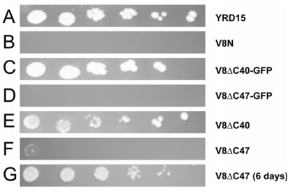
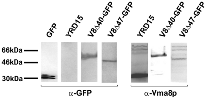
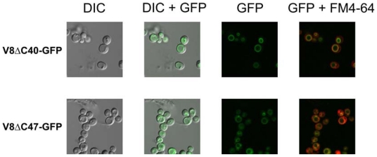
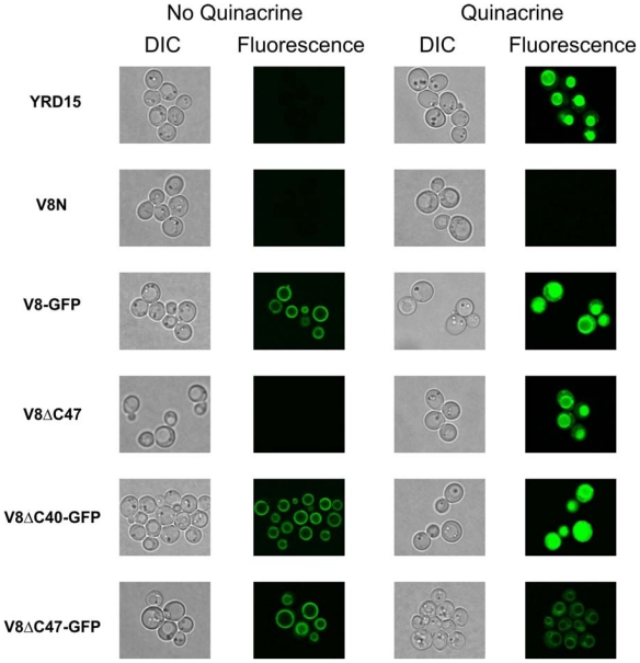
The vacuolar ATPase (V-ATPase) complex of yeast (Saccharomyces cerevisiae) is comprised of two sectors, V(1) (catalytic) and V(O) (proton transfer). The hexameric (A(3)B(3)) cylinder of V(1) has a central cavity that must accommodate at least part of the rotary stalk of V-ATPase, a key component of which is subunit D (Vma8p). Recent electron microscopy (EM) data for the prokaryote V-ATPase complex (Thermus thermophilus) suggest that subunit D penetrates deeply into the central cavity. The functional counterpart of subunit D in mitochondrial F(1)F(O)-ATP synthase, subunit γ, occupies almost the entire length of the central cavity. To test whether the structure of yeast Vma8p mirrors that of subunit γ, we probed the location of the C-terminus of Vma8p by attachment of a large protein adduct, green fluorescent protein (GFP). We found that truncated Vma8p proteins lacking up to 40 C-terminal residues fused to GFP can be incorporated into functional V-ATPase complexes, and are able to support cell growth under alkaline conditions. We conclude that large protein adducts can be accommodated at the top of the central cavity of V(1) without compromising V-ATPase function, arguing for structural flexibility of the V(1) sector.
Keywords: V-ATPase; Vma8p; yeast.
Figures
Similar articles
-
Crystal and NMR structures give insights into the role and dynamics of subunit F of the eukaryotic V-ATPase from Saccharomyces cerevisiae.J Biol Chem. 2013 Apr 26;288(17):11930-9. doi: 10.1074/jbc.M113.461533. Epub 2013 Mar 8. J Biol Chem. 2013. PMID: 23476018 Free PMC article.
-
Origin of asymmetry at the intersubunit interfaces of V1-ATPase from Thermus thermophilus.J Mol Biol. 2013 Aug 9;425(15):2699-708. doi: 10.1016/j.jmb.2013.04.022. Epub 2013 Apr 29. J Mol Biol. 2013. PMID: 23639357
-
Identification of a domain in the V0 subunit d that is critical for coupling of the yeast vacuolar proton-translocating ATPase.J Biol Chem. 2006 Oct 6;281(40):30001-14. doi: 10.1074/jbc.M605006200. Epub 2006 Aug 4. J Biol Chem. 2006. PMID: 16891312
-
Rotation, structure, and classification of prokaryotic V-ATPase.J Bioenerg Biomembr. 2005 Dec;37(6):405-10. doi: 10.1007/s10863-005-9480-1. J Bioenerg Biomembr. 2005. PMID: 16691473 Review.
-
Structural features and nucleotide-binding capability of the C subunit are integral to the regulation of the eukaryotic V1Vo ATPases.Biochem Soc Trans. 2005 Aug;33(Pt 4):883-5. doi: 10.1042/BST0330883. Biochem Soc Trans. 2005. PMID: 16042619 Review.
Cited by
-
Cryo-EM structure of the homohexameric T3SS ATPase-central stalk complex reveals rotary ATPase-like asymmetry.Nat Commun. 2019 Feb 7;10(1):626. doi: 10.1038/s41467-019-08477-7. Nat Commun. 2019. PMID: 30733444 Free PMC article.
References
-
- Arata Y, Nishi T, Kawasaki-Nishi S, Shao E, Wilkens S, Forgac M. Structure, subunit function and regulation of the coated vesicle and yeast vacuolar (H(+))-ATPases. Biochim. Biophys. Acta. 2002;1555:71–74. - PubMed
-
- Forgac M. Vacuolar ATPases: Rotary proton pumps in physiology and pathophysiology. Nat. Rev. Mol. Cell Biol. 2007;8:917–929. - PubMed
-
- Graham LA, Powell B, Stevens TH. Composition and assembly of the yeast vacuolar H(+)-ATPase complex. J. Exp. Biol. 2000;203:61–70. - PubMed
-
- Venzke D, Domgall I, Kocher T, Fethiere J, Fischer S, Bottcher B. Elucidation of the stator organization in the V-ATPase of Neurospora crassa. J. Mol. Biol. 2005;349:659–669. - PubMed
Publication types
MeSH terms
Substances
LinkOut - more resources
Full Text Sources
Molecular Biology Databases
