Review
. 2011 Aug;1(2):110-7.
doi: 10.1016/j.coviro.2011.05.019.

Near-atomic-resolution cryo-EM for molecular virology

Affiliations
Review

Near-atomic-resolution cryo-EM for molecular virology

Corey F Hryc et al. Curr Opin Virol. 2011 Aug.

Abstract

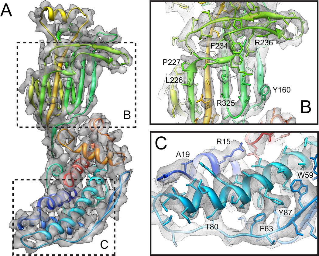
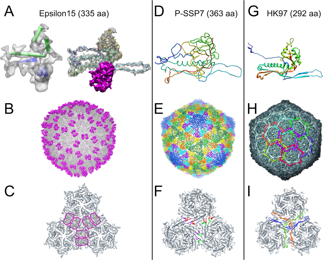
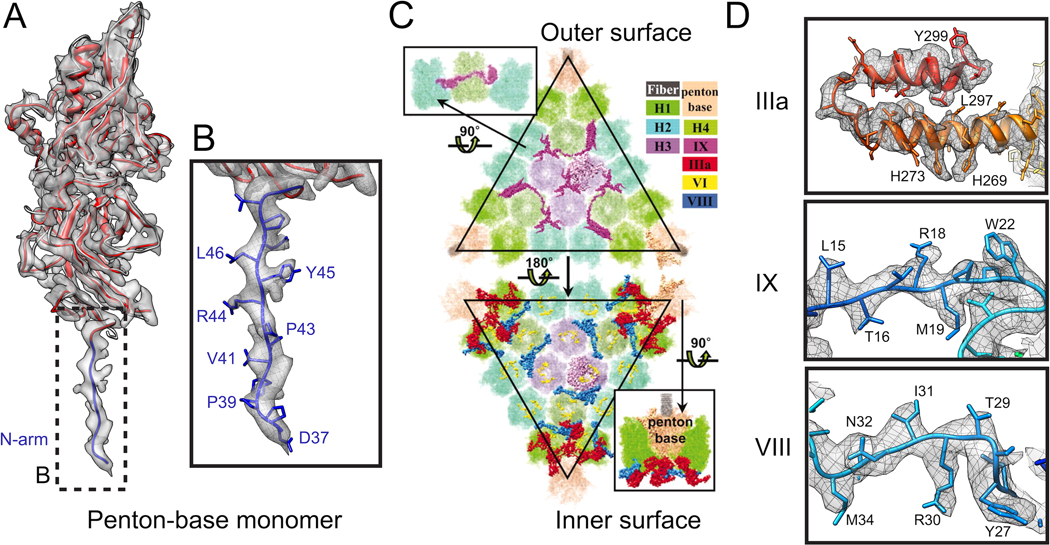
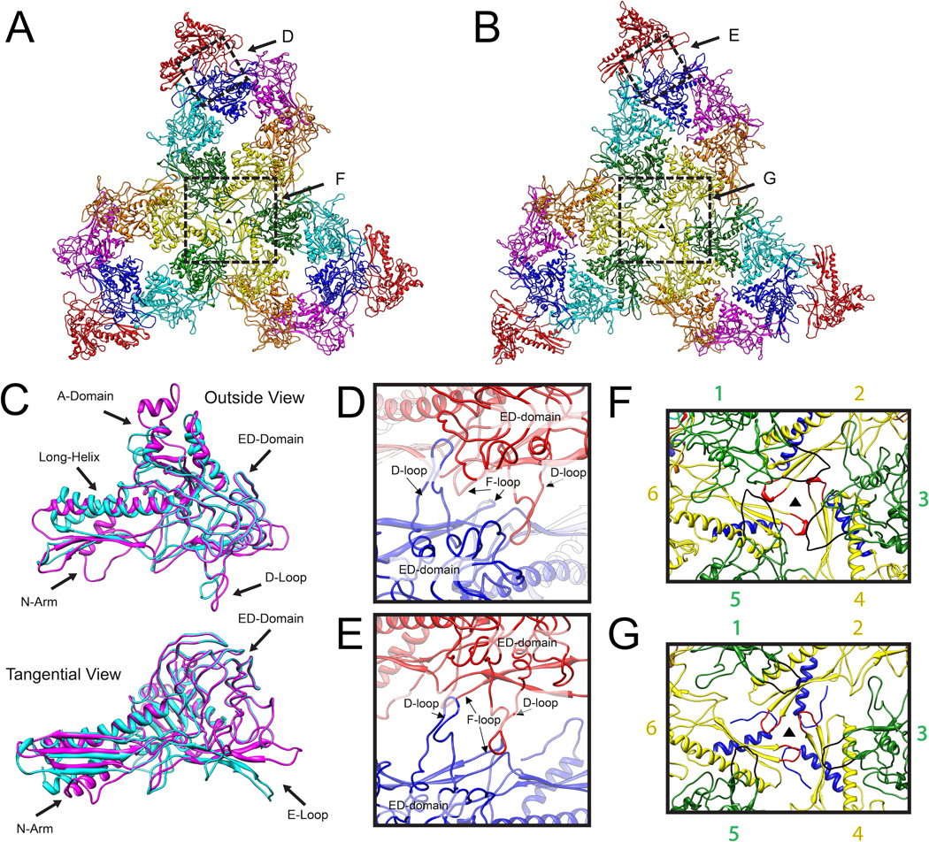
Electron cryo-microscopy (cryo-EM) is a technique in structural biology that is widely used to solve the three-dimensional structures of macromolecular assemblies, close to their biological and solution conditions. Recent improvements in cryo-EM and single-particle reconstruction methodologies have led to the determination of several virus structures at near-atomic resolution (3.3 - 4.6 Å). These cryo-EM structures not only resolve the Cα backbones and side-chain densities of viral capsid proteins, but also suggest functional roles that the protein domains and some key amino acid residues play. This paper reviews the recent advances in near-atomic-resolution cryo-EM for probing the mechanisms of virus assembly and morphogenesis.


Figures

Figure 1
Figure 1
Rotavirus VP6 structure generated by cryo-EM and single-particle reconstruction method (EMDB ID: 1461) [15]. (A) cryo-EM map of VP6 shown at 3.8 Å resolution along with the corresponding crystal structure (PDB ID: 1QHD) [43]. At this resolution, α-helices and β-sheets are well defined, and bulky side-chains are seen protruding from the density. (B) Individual stands in the β-sheet region begin to separate and the loops connecting the strands are defined. Docking the crystal structure into the density map reveals residues that correspond to the bulky side-chains. (C) Helix (amino acids 75-100) showing helical grooves along with bulky densities where individual residues from the X-ray model fit into the density. In addition, the loop region (residues 61-74; shown at the bottom) between SSEs has well defined density trace and bulky side-chain densities that match the corresponding X-ray structure.
Figure 2
Figure 2
New proteins and individual folds are discovered with high-resolution structures. (A - Left) At 9 Å resolution (EMDB ID: 1176) [19] globular domains are revealed for the structure and larger SSEs can be identified in the map. SSEs were identified for ε15 revealing α-helices (green cylinders) and β-sheets (blue planes). (A - Right) Improving the 9 Å map to 4.5 Å in resolution (EMDB ID: 5003) [9], reveals the Cα trace for the capsid protein (shown inside grey density) (PDB ID: 3C5B), as well as density for the newly resolved auxiliary protein gp10 (magenta density). This density was previously thought to be part of the capsid protein. (B) The complete ε15 density map generated with the gp7 models shown with gp10 colored in magenta. (C) The location of gp10 is circled in magenta, and acts as a staple across the two fold, assisting in capsid stability. (D) P-SSP7 capsid protein model with 363 amino acids resolved (PDB ID: 2XD8) [10]. (E) The complete P-SSP7 capsid built with the generated models revealing extending loops and interacting subunits. (F) Interacting P-domains highlighted in red, purple, and green for individual asymmetric units. (G) HK-97 capsid protein model with 292 amino acids resolved (PDB ID: 1OHG) [20]. (H) The complete HK-97 capsid protein built with generated models, revealing the complexity of the chainmail interactions, used for stabilizing the capsid. (I) Chainmail interactions located at the 3-fold, highlighted in blue, orange, and green for individual asymmetric unit.
Figure 3
Figure 3
The cryo-EM 3.6 Å structure of adenovirus reveals new regions as well as a network of interactions [11]. (A) The cryo-EM model (PDB ID: 3IYN) of the penton-based monomer (red) is similar to the X-ray structure (PDB ID: 1VSZ) (gray) [25], however, the cryo-EM structure reveals 14 unseen residues (shown in blue). (B) These 14 newly resolved residues (amino acids 37-51) are part of the N-arm and have multiple bulky side-chains that fit well in the cryo-EM density. (C) Inside view and outside view of adenovirus revealing minor and major proteins and their interactions resolved by cryo-EM (EMDB ID: 5172). (D) Selected regions of the three minor proteins (IIIa, IX, and VIII) reveal side-chain densities, thus allowing for specific interactions to be discovered.
Figure 4
Figure 4
Conformational changes identified by resolving the cryo-EM structures of the P22 procapsid and the virion to near-atomic resolution [5]. (A) Three procapsid asymmetric units are shown to reveal interactions, in addition to spatial arrangements of individual subunits. Skewing of the hexamer subunits results in a large hexamer opening which allows scaffolding protein to exit the virion upon DNA packaging (PDB ID: 2XYY). (B) Three virion asymmetric units are shown to reveal interactions, in addition to spatial arrangements of individual subunits (PDB ID: 2XYZ). (C) Subunit differences between the procapsid (cyan) and virion (magenta) are shown with an outside view (top) as well as a tangential view (bottom). Multiple domains such as the A-domain, long-helix, D-loop, N-arm and E-loop undergo large conformational changes upon capsid maturation. (D) The 2-fold axis of the procapsid shows two neighboring subunits interacting through electrostatic charges. The D-loop of both subunits sits in a pocket of opposite charged residues beside the adjacent ED-domain. (E) Upon maturation, the D-loops pull out of the pocket, become closer and interact to maintain capsid stability. (F) The 3-fold axis reveals 6 subunits (numbered on sides) that play a role in capsid stabilization for the procapsid. This location contains interactions between the P-domains (shown in red) and E-loops (shown in black). (G) The interactions at the 3-fold axis for the virion. The P-domains (red) still remain vital in capsid stabilization, interacting with N-arm regions (shown in blue) across capsomeres, while the E-loops are pulled out from the 3-fold location to interact with the long-helix and β-sheet within capsomeres (reproduced from [5]).

Similar articles

Cited by

References

    1. Harrison SC. Principles of virus structure. Fields virology. 2001;1:53–85.
    1. Adrian M, Dubochet J, Lepault J, McDowall AW. Cryo-electron microscopy of viruses. Nature. 1984;308:32–36. - PubMed
    1. Glaeser RM. Electron crystallography of biological macromolecules. USA: Oxford University Press; 2007.
    1. De Rosier DJ, Klug A. Reconstruction of three dimensional structures from electron micrographs. Nature. 1968;217:130–134. - PubMed
    1. Chen DH, Baker ML, Hryc CF, Dimaio F, Jakana J, Wu W, Dougherty M, Haase-Pettingell C, Schmid MF, et al. Structural basis for scaffolding-mediated assembly and maturation of a dsDNA virus. Proc Natl Acad Sci U S A. 2011;108:1355–1360. Cryo-EM was used to resolve two morphogenetic states of the P22 phage at near-atomic resolution, revealing detailed conformational changes upon maturation. A virus assembly mechanism was proposed based on the interactions among the capsid protein, the newly identified scaffolding proteins, and the portal complex. A similar viral mechanism may be found in some dsDNA viruses.

Publication types

Substances

LinkOut - more resources