. 2011 Sep;18(9):832-41.
doi: 10.1177/1933719111398501.

AMG 479, a novel IGF-1-R antibody, inhibits endometrial cancer cell proliferation through disruption of the PI3K/Akt and MAPK pathways

Affiliations

AMG 479, a novel IGF-1-R antibody, inhibits endometrial cancer cell proliferation through disruption of the PI3K/Akt and MAPK pathways

Albert Mendivil et al. Reprod Sci. 2011 Sep.

Abstract

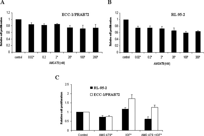
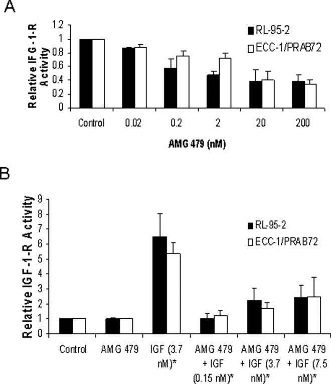
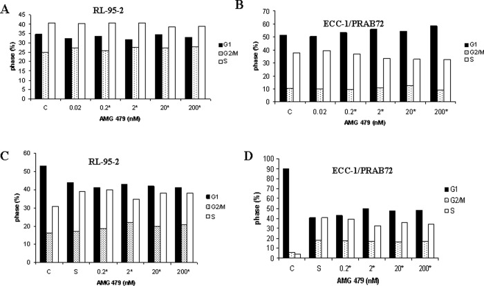
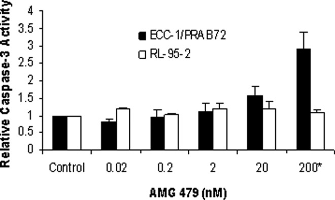
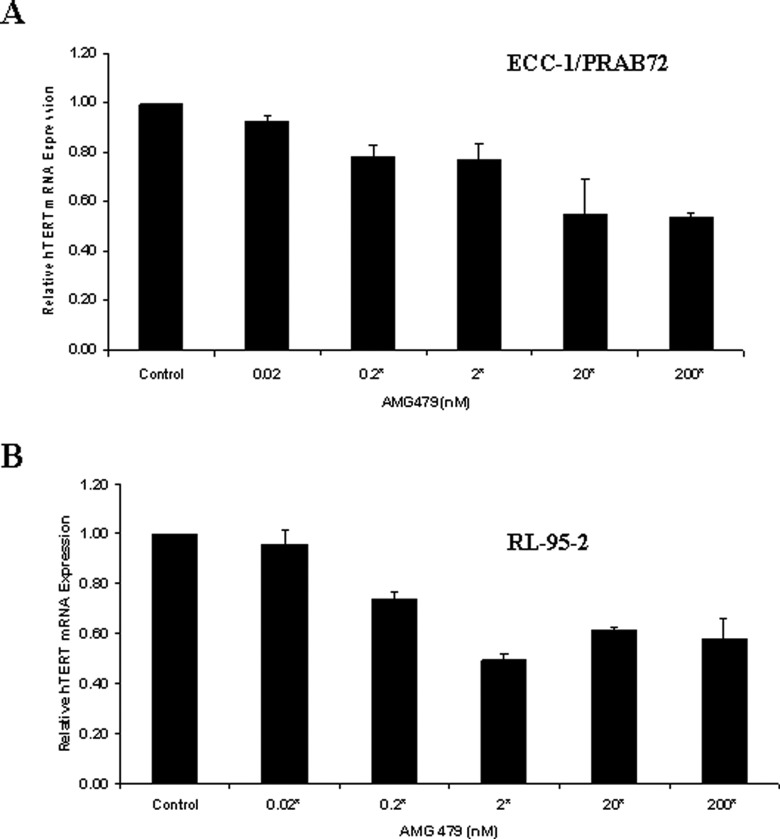
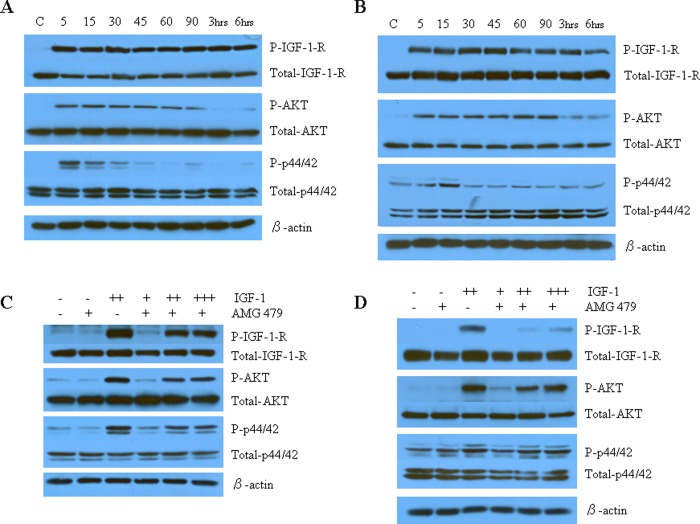
Our goal was to evaluate the therapeutic potential of a novel antibody to the insulin growth factor-1 receptor (IGF-1-R; AMG 479) in endometrial cancer cells. The endometrial cancer cell lines, ECC-1/PRAB72 and RL-95-2, were used. Treatment with AMG 479 (0.02-200 nmol/L) resulted in inhibition of cell proliferation at 72 to 120 hours. Insulin growth factor-1 (0.15-7.5 nmol/L) stimulated growth in both cell lines (range of 15%-42%, P = .0025-.0445), which could be blocked by pretreatment with AMG 479 (mean of 29% for ECC-1/PRAB72, P = .006-.007; mean of 36% for RL-95-2, P = .0002-.0045). AMG 479 suppressed IGF-1-R kinase activity in a dose-dependent manner. Cells treated with AMG 479 underwent either G1 (ECC-1/PRAB72) or G2 (RL-95-2) arrest. AMG 479 decreased human telomerase reverse transcriptase (hTERT) mRNA expression in both endometrial cancer cell lines. Treatment with AMG 479 rapidly blocked IGF-1-induced phosphorylation of IFG-1-R, Akt, and p44/42. Thus, manipulation of the IGF-1-R pathway may serve as a promising therapeutic strategy for the treatment of endometrial cancer.


Conflict of interest statement

The authors declared no conflicts of interest with respect to the authorship and/or publication of this article.

Figures

Figure 1.
Figure 1.
Effect of AMG 479 on proliferation of endometrial cancer cells. The ECC-1/PRAB72 (A) and RL-95-2 (B) cell lines were cultured in the presence of varying concentrations of AMG 479 for 5 days. AMG 479 inhibited proliferation in both of these cell lines. Subsequently, both endometrial cancer cell lines were pretreated with AMG 479 (6.6 nmol/L) for 6 hours and then exposed to insulin growth factor-1 (IGF-1; 3.7 nmol/L) for 72 hours. AMG 479 blocked IGF-1-induced cell proliferation in both of these cell lines (C). Relative growth of cells was determined by MTT assay. The results are shown as the mean ± SE of triplicate samples and are representative of 3 independent experiments (* Indicates statistically significant difference.).
Figure 2.
Figure 2.
The effects of AMG 479 on insulin growth factor-1 receptor (IGF-1-R) activity. RL-95-2 and ECC-1/PRAB72 cells were starved overnight and then treated with 5% fetal bovine serum (FBS) and varying concentrations of AMG 479 alone for 60 minutes (A), or treated with AMG (2 nmol/L) in combination with IGF-1 for 15 minutes (B). The phosphotyrosine levels (Tyr 1131) of the activated IGF-1-R were measured by enzyme-linked immunosorbent assay (ELISA). Treatment with AMG 479 alone significantly reduced IGF-1-R activity in a dose-dependent manner in both of the endometrial cancer cell lines (A). As expected, cells treated with IGF-1 alone (3.7 nmol/L) for 15 minutes demonstrated a dramatic increase in IGF-1-R kinase activity (B). AMG 479 (2 nmol/L) was able to potently block IGF-1 (0.15-7.5 nmol/L) stimulated autophosphorylation of the IGF-1-R in both endometrial cancer cell lines (B). These results are representative of two2 independent experiments (* Indicates statistically significant difference.).
Figure 3.
Figure 3.
Induction of cell-cycle arrest by AMG 479. RL-95-2 and ECC-1/PRAB72 cells were cultured for 24 hours and treated with AMG 479 at varying concentrations in 5% fetal bovine serum (FBS) for 36 hours (A and B) or starved overnight and then stimulated with 15% serum and AMG 479 at the noted concentrations for 36 hours (C and D). Cell-cycle analysis was performed by flow cytometry. AMG 479 inhibited cell-cycle progression by arrest in G1 phase in the ECC-1/PRAB72 cells, and G2/M phase in the RL-95-2 cells. Results shown are representative of 1 of the 3 independent experiments. Statistical significant results are indicated by a *, as determined using the Student t test.
Figure 4.
Figure 4.
The effects of AMG 479 on apoptosis. The RL-95-2 and ECC-1/PRAB72 cells were cultured for 24 hours and then treated with AMG 479 at the indicated concentrations for an additional 24 hours. Apoptosis was assessed using an antibody to caspase-3. AMG 479 increased apoptosis in the ECC-1/PRAB72 cell line but only at high doses of treatment (200 nmol/L). Lower doses of AMG 479 had little effect on caspase-3 activity in this cell line. For the RL-95-2 cell line, there was no significant increase in caspase-3 activity at any dose of AMG 479. The results are shown as the mean ± SD and are representative of 2 independent experiments (* indicates statistically significant difference.).
Figure 5.
Figure 5.
AMG 479 decreased human telomerase reverse transcriptase (hTERT) mRNA expression in a dose-dependent manner in (A) ECC-1/PRAB72 and (B) RL-95-2 cells. Both cells were cultured for 24 hours and then treated with the indicated concentrations of AMG 479 for an additional 48 hours. Human telomerase reverse transcriptase expression was determined by real-time reverse transcription polymerase chain reaction (RT-PCR). The results are shown as the mean ± SE of 2 independent experiments (* indicates statistically significant difference.).
Figure 6.
Figure 6.
The effect of AMG 479 on IGF-1-R and its downstream cell signaling targets as assessed by Western immunoblotting. The ECC-1/PRAB72 (A and C) and RL-95-2 (B and D) cell lines were cultured for 24 hours followed by starving for 12 hours. The cell media was replaced with fresh media containing 0.5% stripped fetal bovine serum (FBS) and AMG 479 or IGF-1 as indicated. (A and B) IGF-1 activated IGF-1-R and its signaling pathway. Insulin growth factor-1 (7.5 nmol/L) alone rapidly induced phosphorylation of IGF-1-R in both of the endometrial cancer cell lines. Similarly, treatment with IGF-1 resulted in phosphorylation of both Akt and MAPK (p42/44) after only 5 minutes of exposure. (C and D) AMG 479 blocked the phosphorylation of IGF-1-R, AKT, and p42/44 induced by IGF-1. Serum-starved ECC-1/PRAB72 and RL-95-2 cells were pretreated with AMG 479 (6.6 nmol/L) for 3 hours and subsequently stimulated with different concentrations of IGF-1 for 15 minutes (+, 0.15 nmol/L; ++, 3.7 nmol/L; +++, 7.5 nmol/L). These results are representative of 2 independent experiments. There was little effect on total-Akt, total-p42/p44, or total IGF-1-R expression in either of these experiments.

References

    1. Chia VM, Newcomb PA, Trentham-Dietz A, Hampton JM. Obesity, diabetes, and other factors in relation to survival after endometrial cancer diagnosis. Int J Gynecol Cancer. 2007;17(2):441–446 - PubMed
    1. Cust AE, Kaaks R, Friedenreich C, et al. Plasma adiponectin levels and endometrial cancer risk in pre- and postmenopausal women. J Clin Endocrinol Metab. 2007;92(1):255–263 - PubMed
    1. Friberg E, Mantzoros CS, Wolk A. Diabetes and risk of endometrial cancer: a population-based prospective cohort study. Cancer Epidemiol Biomarkers Prev. 2007;16(2):276–280 - PubMed
    1. Soliman PT, Wu D, Tortolero-Luna G, et al. Association between adiponectin, insulin resistance, and endometrial cancer. Cancer. 2006;106(11):2376–2381 - PubMed
    1. McCampbell AS, Broaddus RR, Loose DS, Davies PJ. Overexpression of the insulin-like growth factor I receptor and activation of the AKT pathway in hyperplastic endometrium. Clin Cancer Res. 2006;12(21):6373–6378 - PubMed

Publication types

MeSH terms

LinkOut - more resources