. 2011 Sep 30;286(39):33729-36.
doi: 10.1074/jbc.M111.291591. Epub 2011 Aug 16.

Mammalian ACSF3 protein is a malonyl-CoA synthetase that supplies the chain extender units for mitochondrial fatty acid synthesis

Affiliations

Mammalian ACSF3 protein is a malonyl-CoA synthetase that supplies the chain extender units for mitochondrial fatty acid synthesis

Andrzej Witkowski et al. J Biol Chem. .

Abstract

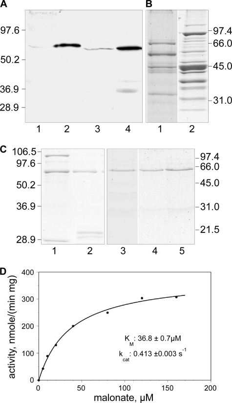
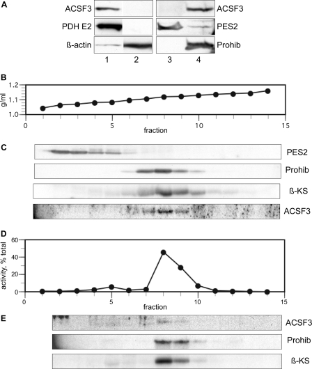
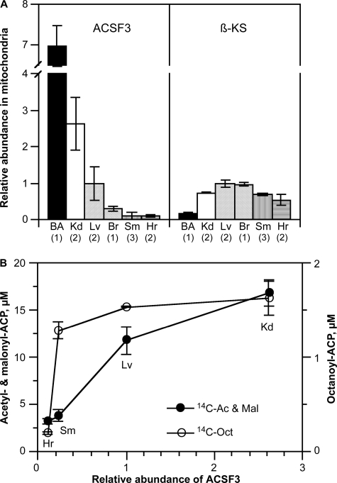
The objective of this study was to identify a source of intramitochondrial malonyl-CoA that could be used for de novo fatty acid synthesis in mammalian mitochondria. Because mammalian mitochondria lack an acetyl-CoA carboxylase capable of generating malonyl-CoA inside mitochondria, the possibility that malonate could act as a precursor was investigated. Although malonyl-CoA synthetases have not been identified previously in animals, interrogation of animal protein sequence databases identified candidates that exhibited sequence similarity to known prokaryotic forms. The human candidate protein ACSF3, which has a predicted N-terminal mitochondrial targeting sequence, was cloned, expressed, and characterized as a 65-kDa acyl-CoA synthetase with extremely high specificity for malonate and methylmalonate. An arginine residue implicated in malonate binding by prokaryotic malonyl-CoA synthetases was found to be positionally conserved in animal ACSF3 enzymes and essential for activity. Subcellular fractionation experiments with HEK293T cells confirmed that human ACSF3 is located exclusively in mitochondria, and RNA interference experiments verified that this enzyme is responsible for most, if not all, of the malonyl-CoA synthetase activity in the mitochondria of these cells. In conclusion, unlike fungi, which have an intramitochondrial acetyl-CoA carboxylase, animals require an alternative source of mitochondrial malonyl-CoA; the mitochondrial ACSF3 enzyme is capable of filling this role by utilizing free malonic acid as substrate.


Figures

FIGURE 1.
FIGURE 1.
Purification and properties of ACSF3. A, Western analysis of bovine and mouse mitochondrial extracts probed with antibody against ACSF3. Lanes 1 and 3, 60 μg of mitochondrial protein from bovine and mouse heart extracts, respectively; lanes 2 and 4, 10 μg of protein from partially purified bovine and mouse preparations, respectively. B, general protein staining of partially purified bovine (lane 1) and mouse (lane 2) ACSF3 preparations. C, General protein staining of recombinant ACSF3 protein preparations. Lane 1, purified GST-ACSF3; lane 2, GST-ACSF3 after treatment with tobacco etch virus protease; lanes 3–5, final purified preparations of wild-type ACSF3 and mutants R354A and R354L, respectively. D, malonyl-CoA synthetase activity of the ACSF3 preparation. Assays were performed at 37 °C for 30 min, and reaction components were separated by HPLC.
FIGURE 2.
FIGURE 2.
Subcellular localization of ACSF3 in HEK293T cells. A, Western analysis of the mitochondrial fraction from 1.2 × 10⁶ HEK293T cells (lane 1), the post-mitochondrial supernatant from 0.7 × 10⁶ cells (lane 2), the microsomal fraction from 5.8 × 10⁶ cells (lane 3), and the mitochondrial fraction from 5.8 × 10⁶ cells (lane 4). Blots were probed with antibodies against ACSF3, the pyruvate dehydrogenase E2 subunit (PDH E2; a mitochondrial marker), prostaglandin E synthase 2 (PES2; a microsomal marker), β-actin (a cytosolic marker), and prohibitin (Prohib; a mitochondrial marker). B–E, subcellular fractionation of HEK293T cell extracts by centrifugation on an iodixanol density gradient. B, iodixanol density. C, Western analysis of gradient fractions after precipitation with methanol/chloroform. Pellets analyzed for ACSF3 were additionally washed with ice-cold acetone. β-KS, β-ketoacyl synthase (a mitochondrial marker). D, malonyl-CoA synthetase activity. Fractions were diluted and recentrifuged at 100,000 × g for 1 h, the pellet was extracted with 1% Triton X-100, and extracts were assayed as described under “Experimental Procedures.” E, Western analysis of 1% Triton X-100 extracts.
FIGURE 3.
FIGURE 3.
Knockdown of malonyl-CoA synthetase in HEK293T cells by RNA interference. A, Western analysis of mitochondrial extracts from HEK293T cells treated with vehicle only (lane 1), 20 nm ACP siRNA (lane 2), or 75 nm ACSF3 siRNA (lane 3). Loading was normalized on the basis of citrate synthase activity (14.5 units/lane). B, digitalized Western blot data. β-KS, β-ketoacyl synthase. C, malonyl-CoA synthetase activity in mitochondrial extracts. The 100% value for malonyl-CoA synthetase activity corresponds to 2.9 pmol/min, normalized for 1 unit of citrate synthase activity. D, analysis of ACP-linked thioesters formed from [2-14C]malonate metabolism by mitochondrial preparations derived from siRNA-treated HEK293T cells. The metabolites detected included malonyl-ACP and its decarboxylation product, acetyl-ACP, and the two major products of fatty acid synthesis, octanoyl-ACP and hexanoyl-ACP. The 100% value for the [14C]acetyl-ACP plus [14C]malonyl-ACP (14C Acet & Mal-ACP) pool size was 6.4 μm; acetyl-ACP accounted for 35 ± 7% of the total pool. The 100% value for [14C]hexanoyl-ACP was 0.51 μm, and that for [14C]octanoyl-ACP was 0.62 μm, both expressed in terms of malonyl moieties. The data represent means of at least two experiments, with probability values of Student's t test, when <0.1, shown.
FIGURE 4.
FIGURE 4.
Relative abundance of ACSF3 and β-ketoacyl synthase in mitochondria isolated from mouse tissues. A, proteins were detected by Western blotting, and the data were digitized. Numbers in parentheses indicate the number of different mitochondrial preparations analyzed. BA, brown adipose tissue; Kd, kidney; Lv, liver; Br, brain; Sm, skeletal muscle; Hr, heart. B, relationship between products formed from malonate in mitochondrial preparations from different tissues and the relative abundance of ACSF3. Mitochondria used in these experiments were purified on a discontinuous iodixanol gradient as described under “Experimental Procedures.” 14C Ac & Mal, [14C]acetyl-ACP plus [14C]malonyl-ACP; 14C-Oct, [14C]octanoyl-ACP.

Similar articles

Cited by

References

    1. Autio K. J., Kastaniotis A. J., Pospiech H., Miinalainen I. J., Schonauer M. S., Dieckmann C. L., Hiltunen J. K. (2008) FASEB J. 22, 569–578 - PubMed
    1. Chen Z., Kastaniotis A. J., Miinalainen I. J., Rajaram V., Wierenga R. K., Hiltunen J. K. (2009) FASEB J. 23, 3682–3691 - PubMed
    1. Miinalainen I. J., Chen Z. J., Torkko J. M., Pirilä P. L., Sormunen R. T., Bergmann U., Qin Y. M., Hiltunen J. K. (2003) J. Biol. Chem. 278, 20154–20161 - PubMed
    1. Zhang L., Joshi A. K., Hofmann J., Schweizer E., Smith S. (2005) J. Biol. Chem. 280, 12422–12429 - PubMed
    1. Zhang L., Joshi A. K., Smith S. (2003) J. Biol. Chem. 278, 40067–40074 - PubMed

Publication types

MeSH terms