. 2011 Oct 21;286(42):36238-47.
doi: 10.1074/jbc.M111.251900. Epub 2011 Aug 16.

Evidence that diacylglycerol acyltransferase 1 (DGAT1) has dual membrane topology in the endoplasmic reticulum of HepG2 cells

Affiliations

Evidence that diacylglycerol acyltransferase 1 (DGAT1) has dual membrane topology in the endoplasmic reticulum of HepG2 cells

Haja R Wurie et al. J Biol Chem. .

Abstract

Triacylglycerol (TAG) synthesis and secretion are important functions of the liver that have major impacts on health, as overaccumulation of TAG within the liver (steatosis) or hypersecretion of TAG within very low density lipoproteins (VLDL) both have deleterious metabolic consequences. Two diacylglycerol acyltransferases (DGATs 1 and 2) can catalyze the final step in the synthesis of TAG from diacylglycerol, which has been suggested to play an important role in the transfer of the glyceride moiety across the endoplasmic reticular membrane for (re)synthesis of TAG on the lumenal aspect of the endoplasmic reticular (ER) membrane (Owen, M., Corstorphine, C. C., and Zammit, V. A. (1997) Biochem. J. 323, 17-21). Recent topographical studies suggested that the oligomeric enzyme DGAT1 is exclusively lumen facing (latent) in the ER membrane. By contrast, in the present study, using two specific inhibitors of human DGAT1, we present evidence that DGAT1 has a dual topology within the ER of HepG2 cells, with approximately equal DGAT1 activities exposed on the cytosolic and lumenal aspects of the ER membrane. This was confirmed by the observation of the loss of both overt (partial) and latent (total) DGAT activity in microsomes prepared from livers of Dgat1(-/-) mice. Conformational differences between DGAT1 molecules having the different topologies were indicated by the markedly disparate sensitivities of the overt DGAT1 to one of the inhibitors. These data suggest that DGAT1 belongs to the family of oligomeric membrane proteins that adopt a dual membrane topology.


Figures

FIGURE 1.
FIGURE 1.
Chemical structures of the unrelated compounds used to inhibit human DGAT1. A, the structure of inhibitor compound A (iA), 2-((1s,4s)-4-(4-(4-amino-7,7-dimethyl-7H-pyrimido[4,5-b][1,4]oxazin-6-yl)phenyl)cyclohexyl)acetic acid; and B, the structure of compound B (iB), cis-4-{3-fluoro-4-[({5-[(4-fluorophenyl)amino]-1,3,4-oxadiazol-2-yl}carbonyl)amino]phenoxy}cyclohexanecarboxylic acid.
FIGURE 2.
FIGURE 2.
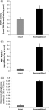
DGAT and marker enzyme activities in intact and alamethacin-permeabilized microsomes from rat liver. Rat liver microsomal fractions were prepared as described under “Experimental Procedures” and incubated on ice either with DMSO carrier (intact, gray bars) or with 20 μg of alamethacin/ml in DMSO (permeabilized, black bars) for 30 min prior to assay of DGAT, (panel i), acyl-CoA ethanol acyltransferase (panel ii), or mannose-6-phosphatase (panel iii) activities at 37 °C. Assays were performed in duplicates. Values are mean ± S.E. for 4 separate preparations.
FIGURE 3.
FIGURE 3.
Dependence of the permeabilization of the plasma and endoplasmic reticular membranes by digitonin treatment of cultured hepatocytes: HepG2 cells (panel i) and McA-RH7777 cells (panel ii). Cells were cultured as described under “Experimental Procedures.” They were detached from the dishes by mild trypsinization followed by treatment with the indicated concentrations of digitonin for 30 min on ice. They were then sedimented. For the assay of glycerol 3-phosphate and lactate dehydrogenase release (markers of permeabilization of the plasma membrane to small and large molecules, respectively) the amounts released into the supernatant were measured (black and gray bars, respectively). For the exposure of mannose-6-phosphatase activity (a nonreleasable marker of permeabilization of the ER membrane) the pellets were resuspended and assayed for activity (white bars) as described under “Experimental Procedures.” Values are expressed as a percentage of the total marker released/exposed by treatment of the cells with 1% Triton, and are mean ± S.E. for 4 separate determinations.
FIGURE 4.
FIGURE 4.
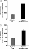
Effects of differential permeabilization of the plasma membrane and the additional permeabilization of the endoplasmic reticulum in whole cells on DGAT activity. The cells maintained in culture were McA-RH7777 cells (panel i) and HepG2 cells (panel ii); they were detached by light trypsinization and treated with digitonin (30 μg/ml) alone (gray bars) or, after washing, for a further period (30 min on ice) in the presence of 20 μg of alamethacin/ml (black bars). They were then assayed for DGAT activity to measure overt and total DGAT activity, respectively. Values are mean ± S.E. for 4 separate cell preparations.
FIGURE 5.
FIGURE 5.
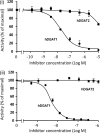
Dose-response curves for the inhibition of human DGAT1 and DGAT2 expressed in insect cell membranes. The human DGATs (hDGAT1 and hDGAT2) were expressed in insect cells using Baculovirus (see “Experimental Procedures”). The effects of increasing concentrations of inhibitors iA and iB (panels i and ii, respectively) on the DGAT1 (●) and DGAT2 (■) activity associated with the membranes were quantified. Values are mean ± S.E. Dose-response curves and IC50 values were generated from three separate determinations, and IC50 values were computed using Origin software and were 38.3 and 3.4 nm for iA and iB, respectively.
FIGURE 6.
FIGURE 6.
DGAT1 inhibitors decrease cellular triglyceride synthesis and secretion in intact HepG2 cells but do not permeabilize the endoplasmic reticular membrane. HepG2 cells were cultured and harvested as described under “Experimental Procedures.” In panel i the percent inhibition (relative to controls to which only carrier DMSO was added) of total cellular DGAT activity (black bars) and incorporation of [3H]glycerol into cellular (gray bars) or secreted triacylglycerol (white bars) was quantified. In panel ii, assays for AEAT activity were performed on cells in the absence (DMSO carrier only) or after treatment with iA (1.5 μm) or iB (240 nm) of either intact cells (white bars) or cells that had their plasma membrane permeabilized by treatment with 30 μg of digitonin/ml alone (gray bars) or additionally cells that had their ER membrane permeabilized by treatment with 20 μg of alamethacin/ml (black bars), as indicated on the x axis legend. For each condition, assays were performed after a further period (30 min) of incubation of the cells with either of the two inhibitor compounds, iA (1.5 μm) or iB (240 nm). The concentration of DMSO in the assay was kept constant at 0.6% vol. Values are mean ± S.E. for 3 separate determinations.
FIGURE 7.
FIGURE 7.
Differential inhibition of overt and latent DGAT1 activities in HepG2 cells by the two inhibitors. HepG2 cells were cultured and harvested as described under “Experimental Procedures.” In panel i, cells were permeabilized with 30 μg of digitonin/ml as described under “Experimental Procedures” (gray bars) and, additionally, with alamethacin (black bars). They were then assayed for overt and total DGAT activity in the absence or presence of either iA (150 nm) or iB (24 nm). In panels ii and iii, the dose-dependent effects of iA and iB on overt and latent DGAT activities, respectively, are shown. Latent DGAT activity (■) was calculated as the difference between total and overt DGAT activities. Values are mean ± S.E. for 3 separate cell preparations.
FIGURE 8.
FIGURE 8.
Overt and total DGAT activities in microsomes prepared from livers of wild-type and Dgat1−/− mice. Microsomal fractions were prepared from the livers of wild-type (gray bars) and Dgat1−/− mice (black bars). They were incubated on ice for 30 min in the absence (Intact) or presence (Permeabilized) of alamethacin (20 μg/ml) and assayed for overt and total DGAT activity, respectively, as described under “Experimental Procedures.” Values are mean ± S.E. for 3 separate determinations.

Similar articles

Cited by

References

    1. Gotto A. M., Jr. (1998) Am. J. Cardiol. 82, 22Q–25Q - PubMed
    1. Zammit V. A., Waterman I. J., Topping D., McKay G. (2001) J. Nutr. 131, 2074–2077 - PubMed
    1. Erion D. M., Shulman G. I. (2010) Nat. Med. 16, 400–402 - PMC - PubMed
    1. Cases S., Smith S. J., Zheng Y. W., Myers H. M., Lear S. R., Sande E., Novak S., Collins C., Welch C. B., Lusis A. J., Erickson S. K., Farese R. V., Jr. (1998) Proc. Natl. Acad. Sci. U.S.A. 95, 13018–13023 - PMC - PubMed
    1. Cases S., Stone S. J., Zhou P., Yen E., Tow B., Lardizabal K. D., Voelker T., Farese R. V., Jr. (2001) J. Biol. Chem. 276, 38870–38876 - PubMed

Publication types

LinkOut - more resources