. 2011 Sep;134(Pt 9):2772-86.
doi: 10.1093/brain/awr197. Epub 2011 Aug 16.

Epstein-Barr virus in the multiple sclerosis brain: a controversial issue--report on a focused workshop held in the Centre for Brain Research of the Medical University of Vienna, Austria

Collaborators, Affiliations

Epstein-Barr virus in the multiple sclerosis brain: a controversial issue--report on a focused workshop held in the Centre for Brain Research of the Medical University of Vienna, Austria

Hans Lassmann et al. Brain. 2011 Sep.

Abstract

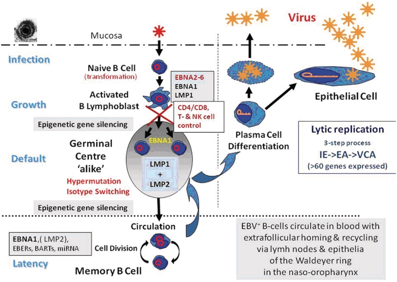
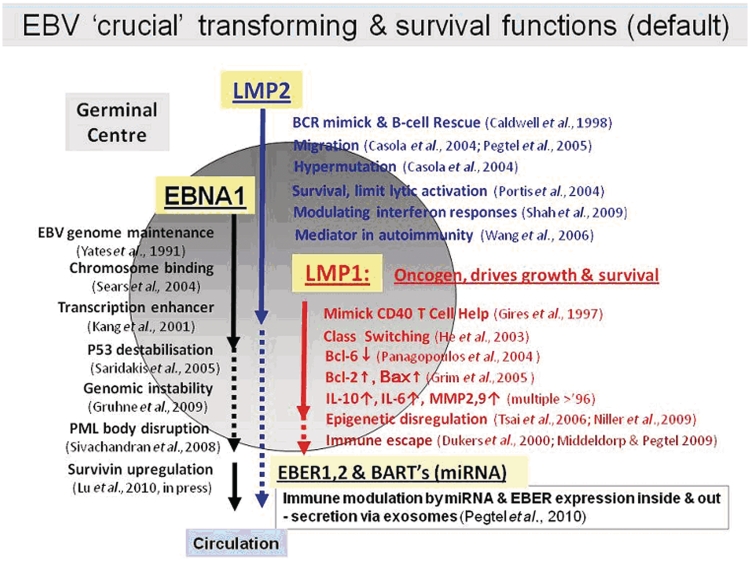
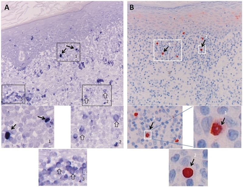
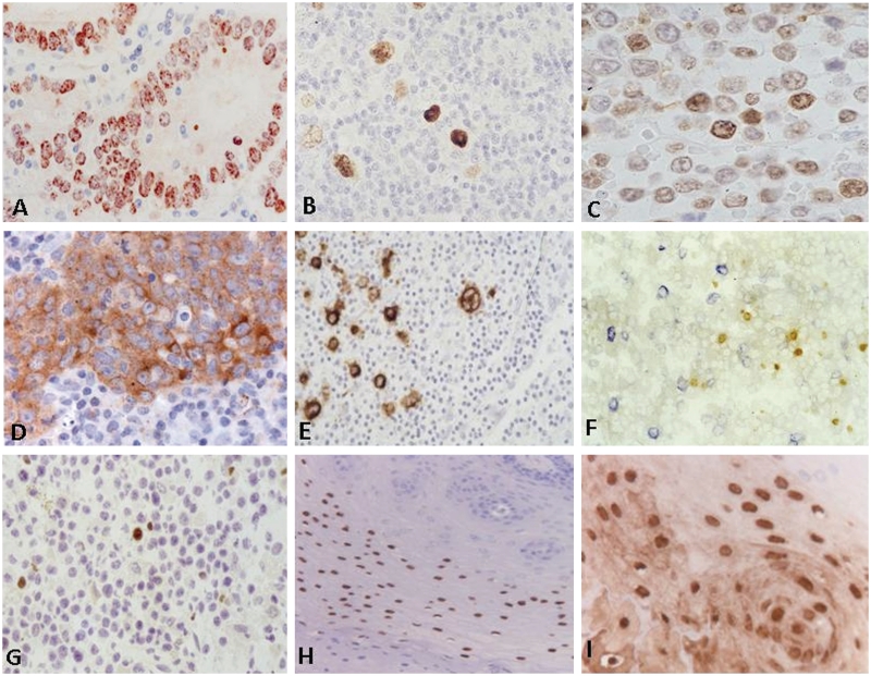
Recent epidemiological and immunological studies provide evidence for an association between Epstein-Barr virus infection and multiple sclerosis, suggesting a role of Epstein-Barr virus infection in disease induction and pathogenesis. A key question in this context is whether Epstein-Barr virus-infected B lymphocytes are present within the central nervous system and the lesions of patients with multiple sclerosis. Previous studies on this topic provided highly controversial results, showing Epstein-Barr virus reactivity in B cells in the vast majority of multiple sclerosis cases and lesions, or only exceptional Epstein-Barr virus-positive B cells in rare cases. In an attempt to explain the reasons for these divergent results, a workshop was organized under the umbrella of the European Union FP6 NeuroproMiSe project, the outcome of which is presented here. This report summarizes the current knowledge of Epstein-Barr virus biology and shows that Epstein-Barr virus infection is highly complex. There are still major controversies, how to unequivocally identify Epstein-Barr virus infection in pathological tissues, particularly in situations other than Epstein-Barr virus-driven lymphomas or acute Epstein-Barr virus infections. It further highlights that unequivocal proof of Epstein-Barr virus infection in multiple sclerosis lesions is still lacking, due to issues related to the sensitivity and specificity of the detection methods.


Figures

Figure 1
Figure 1
Schematic presentation of EBV infection and persistence in vivo. EA = early antigen; IE = immediate early; VCA = viral capsid antigen.
Figure 2
Figure 2
EBV gene expression mimicking the migration and germinal centre selection process. miRNA = microRNA.
Figure 3
Figure 3
EBER peptide nucleic acid staining pitfalls: comparison of detection by alkaline phosphatase and horseradish peroxidise. EBER expression was studied in an inflammatory epithelial lesion using the identical EBER peptide nucleic acid (PNA) probe labelled with FITC (DAKO Kit) applied under identical standardized tissue preparation and hybridization conditions (as described in the DAKO EBER PNA procedure) with subsequent detection (A) according to the manufacturer's protocol with standard rabbit anti-FITC labelled with alkaline phosphatase and BCIP/NBT developing reagent, or (B) using a modified protocol involving rabbit anti-FITC and Streptavidin–horseradish peroxidase (DAKO) detection and diaminobenzidene as developing reagent. Although both methods allowed detection of EBER-positive nuclei (A1 and B1) as appropriate sign of EBV presence in the tissue (black arrows), the alkaline phosphatase detection generally gave a problematic background staining especially in the cytoplasm of non-EBV-infected lymphoid cells (open arrow in A2 and A3). These cells may be plasma cells as judged by the enlarged cytoplasm. Such cytoplasmic staining is regularly observed in infiltrating lymphoid cells with plasma cell appearance in otherwise EBV negative inflammatory tissues. By using the alternative protocol (B), no such cytoplasmic staining was revealed and generally a very sharp nuclear boundary was produced (B1, B2), with occasionally less precise lining (single cell in B1), suggesting of EBER leakage into the cytoplasm. This should be considered when interpreting published data on EBV involvement in multiple sclerosis with commercial detection kits (Serafini et al., 2007, ; Peferoen et al., 2010).
Figure 4
Figure 4
Detection of EBV antigen expression in human tissues. A variety of EBV antigens can be detected by a characteristic staining pattern using defined monoclonal antibody reagents. EBNA1 expression (detected by mouse monoclonal antibody OT1x) is characterized by a (sometimes punctuated) nuclear staining as revealed in gastric cancer (A), Hodgkin's lymphoma (B) and B cell lymphoproliferative lesions (C). LMP1 expression (detected by mouse monoclonal antibody OT21C) is characterized by a homogeneous cytoplasmic and membraneous staining as revealed in nasopharyngeal carcinoma (D), Hodgkin's lymphoma (E) and blastoid cells in post-transplant lymphoproliferative disease (F, LMP1 stained in blue). Note that the parallel expression of nuclear EBNA2 (detected by rat monoclonal antibody R3) in smaller cells in the same lesion (F, EBNA2 stained in brown), suggesting a heterogeneous proliferative process (Brink et al., 1997). Zebra protein expression (detected by the mouse monoclonal antibody BZ1) is strictly nuclear as revealed by staining of post-transplant lymphoproliferative disease (G) or oral hairy leukoplakia (H). The structural capsid protein VCA-p18 can be abundantly detected in oral hairy leukoplakia as revealed by rat monoclonal antibody OT15E (I).

Similar articles

Cited by

References

    1. Aloisi F, Pujol-Borrell R. Lymphoid neogenesis in chronic inflammatory diseases. Nature Rev. 2006;6:205–17. - PubMed
    1. Aloisi F, Serafini B, Magliozzi R, Howell OW, Reynolds R. Detection of Epstein-Barr virus and B-cell follicles in the multiple sclerosis brain: what you find depends on how and where you look. Brain. 2010;133:e157. - PubMed
    1. Ascherio A, Munger KL. Epstein Barr virus infection and multiple sclerosis. J Neuroimmune Pharmacol. 2010;5:271–7. - PubMed
    1. Bonnet M, Guinebretiere JM, Kremmer E, Grunewald V, Benhamou E, Contesso G, et al. Detection of Epstein-Barr virus in invasive breast cancers. J Natl Cancer Inst. 1999;91:1376–81. - PubMed
    1. Brink AA, Dukers DF, van den Brule AJ, Oudejans JJ, Middeldorp JM, et al. Presence of Epstein-Barr virus latency type III at the single cell level in post-transplantation lymphoproliferative disorders and AIDS related lymphomas. J Clin Pathol. 1997;50:911–8. - PMC - PubMed

Publication types

MeSH terms