. 2011 Oct 7;286(40):35257-66.
doi: 10.1074/jbc.M111.269001. Epub 2011 Aug 16.

Mechanism of AMPA receptor activation by partial agonists: disulfide trapping of closed lobe conformations

Affiliations

Mechanism of AMPA receptor activation by partial agonists: disulfide trapping of closed lobe conformations

Ahmed H Ahmed et al. J Biol Chem. .

Abstract

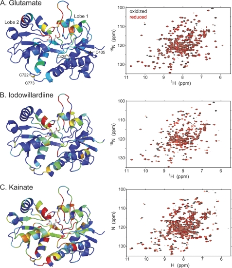
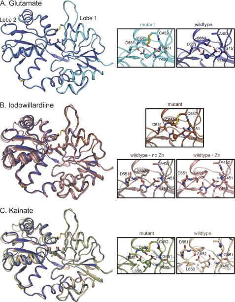
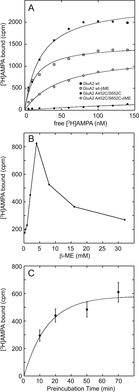
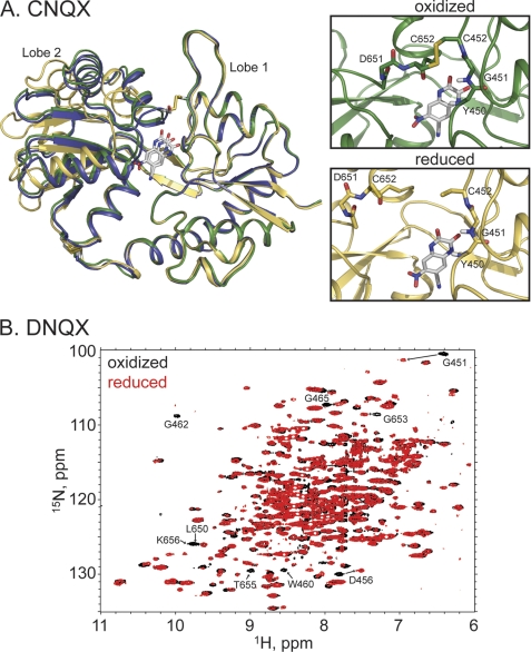
The mechanism by which agonist binding to an ionotropic glutamate receptor leads to channel opening is a central issue in molecular neurobiology. Partial agonists are useful tools for studying the activation mechanism because they produce full channel activation with lower probability than full agonists. Structural transitions that determine the efficacy of partial agonists can provide information on the trigger that begins the channel-opening process. The ligand-binding domain of AMPA receptors is a bilobed structure, and the closure of the lobes is associated with channel activation. One possibility is that partial agonists sterically block full lobe closure but that partial degrees of closure trigger the channel with a lower probability. Alternatively, full lobe closure may be required for activation, and the stability of the fully closed state could determine efficacy with the fully closed state having a lower stability when bound to partial relative to full agonists. Disulfide-trapping experiments demonstrated that even extremely low efficacy ligands such as 6-cyano-7-nitroquinoxaline-2,3-dione can produce a full lobe closure, presumably with low probability. The results are consistent the hypothesis that the efficacy is determined at least in part by the stability of the state in which the lobes are fully closed.


Figures

FIGURE 1.
FIGURE 1.
Comparison of the amide backbone chemical shifts in the oxidized and reduced (DTT) form of A452C/S652C mutant of GluA2 LBD bound to glutamate (A; Protein Data Bank code 3T93), iodowillardiine (B; Protein Data Bank code 3T96), and kainate (C; Protein Data Bank code 3T9H). On the right is shown the 1H,15N-HSQC spectrum, and on the left, a representation of the resonances that have shifted upon reduction of the A452C/S652C disulfide bond is shown. The oxidized form is identical with no additions and in the presence of Cu-phenanthroline, suggesting that the disulfide bond forms spontaneously. The difference in chemical shift between the oxidized and reduced from was quantitated using the formula, chemical shift difference = ((Δ15N)/8)2+(Δ1H)2. Residues were assigned colors with a chemical shift difference between 0 and 5 assigned to a rainbow of colors from blue to red.
FIGURE 2.
FIGURE 2.
Crystal structures of wild type and A452C/S652C mutant GluA2 LBD bound to glutamate (A), iodowillardiine (B), and kainate (C). The right panels show the agonist binding site. A, glutamate. The structure of wild type GluA2 bound to glutamate (Protein Data Bank code 3DP6; B protomer, 24) is shown in dark blue and the A452C/S652C mutant (Protein Data Bank code 3T93) is shown in cyan. The water molecule involved in the Asp-651–Tyr-450 H-bond is shown in the same color as the backbone in all parts of this figure. The Cys-452–Cys-652 disulfide bond is clearly present in the mutant. B, iodowillardiine. The structure bound to glutamate is shown in blue to indicate the orientation of a fully closed lobe. The iodowillardiine-bound structures are shown in shades of brown/pink. Two wild type iodowillardiine-bound structures are shown, one crystallized in the presence of zinc (Protein Data Bank code 1MY4, A protomer; 20) and one in the absence of zinc (Protein Data Bank code 1MQG, A protomer; 12). Both are shown to illustrate the variability seen in different crystal structures. The mutant structure is more closed and the Asp-651-H2O-Tyr-450 and Cys-652–Gly-451 H-bonds are clearly present as is the Cys-452–Cys-652 disulfide bond (Protein Data Bank code 3T96). C, kainate. The glutamate-bound structure is shown in dark blue, and the wild type kainate-bound structure is shown in tan (Protein Data Bank code 1FW0; 3). As seen for iodowillardiine, the A452C/S652C mutant structure (shown in green) is more closed and the Asp-651–H2O–Tyr-450 and Cys-652–Gly-451 H-bonds are clearly present as is the Cys-452–Cys-652 disulfide bond (Protein Data Bank code 3T9H). Note the change in the rotameric state of the side chain of Leu-650.
FIGURE 3.
FIGURE 3.
A, binding of [3H]AMPA to both wild type and the A452C/S652C mutant GluA2 LBD. For the wild type, a decrease in binding was observed in the presence of 5 mm β-metcaptoethanol (apparent decrease in Bmax). In the case of the mutant, binding is only observed when the disulfide bond is reduced by β-metcaptoethanol (β-ME). The KD for binding of [3H]AMPA to the reduced form of the mutant is 2-fold higher than that observed for wild type. B, treatment with β-metcaptoethanol increases binding of the A452C/S652C mutant up to 5 mm, and above that concentration, binding decreases. This may be due to the initial selective reduction of the A452C/S652C followed by the reduction of the Cys-722/Cys-773 disulfide bond. C, the time course for reduction of the disulfide bond shows that it is complete within 60 min in the presence of 5 mm β-metcaptoethanol.
FIGURE 4.
FIGURE 4.
A, crystal structures of the reduced (yellow, Protein Data Bank code 3T9V) and oxidized (green, Protein Data Bank code 3T9U) forms of the A452C/S652C GluA2 LBD mutant bound to CNQX. Also shown is the wild type structure bound to glutamate in blue. The disulfide bond is clearly formed in the oxidized case, with a dramatic change in the relative lobe orientations. B, the 1H,15N-HSQC spectra of the reduced (red) and oxidized (black) forms of the A452C/S652C GluA2 LBD mutant bound to DNQX. Specific chemical shift changes, consistent with the formation of the disulfide bond, are seen.
FIGURE 5.
FIGURE 5.
The 1H,15N-HSQC spectra of the reduced (red) and oxidized (black; Cu-phenanthroline) forms of the A452C/S652C GluA2 LBD mutant bound to UBP282. No chemical shift changes are observed, consistent with the absence of the disulfide bond in the presence of Cu-phenanthroline.
FIGURE 6.
FIGURE 6.
A, full-length wild type, S652C, or A452C/S652C GluA2 was transfected into HEK293 cells, and the response to glutamate was measured using a ratiometric fura-2 imaging. Clear channel activation was observed for wild type and S652C but not for the double mutant. B, immunofluorescent labeling of HEK293 cells transfected with full-length wild type, S652C, or A452C/S652C GluA2. In both cases, the cells were fixed in 2% paraformaldehyde prior to labeling, but the total staining was done in the presence of 0.1% Triton X-100 to allow access of the antibody to the cytoplasm. Surface staining was done in the absence of Triton X-100. The primary antibody recognized an extracellular determinant on GluA2.

References

    1. Dingledine R., Borges K., Bowie D., Traynelis S. F. (1999) Pharmacol. Rev. 51, 7–61 - PubMed
    1. Traynelis S. F., Wollmuth L. P., McBain C. J., Menniti F. S., Vance K. M., Ogden K. K., Hansen K. B., Yuan H., Myers S. J., Dingledine R., Sibley D. (2010) Pharmacol. Rev. 62, 405–496 - PMC - PubMed
    1. Armstrong N., Gouaux E. (2000) Neuron 28, 165–181 - PubMed
    1. Armstrong N., Sun Y., Chen G. Q., Gouaux E. (1998) Nature 395, 913–917 - PubMed
    1. Mayer M. L. (2005) Neuron 45, 539–552 - PubMed

Publication types