. 2011 Aug 16;30(20):4287-98.
doi: 10.1038/emboj.2011.299.

CBP is required for environmental enrichment-induced neurogenesis and cognitive enhancement

Affiliations

CBP is required for environmental enrichment-induced neurogenesis and cognitive enhancement

Jose P Lopez-Atalaya et al. EMBO J. .

Abstract

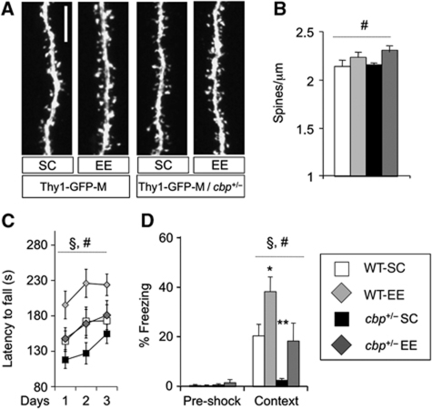
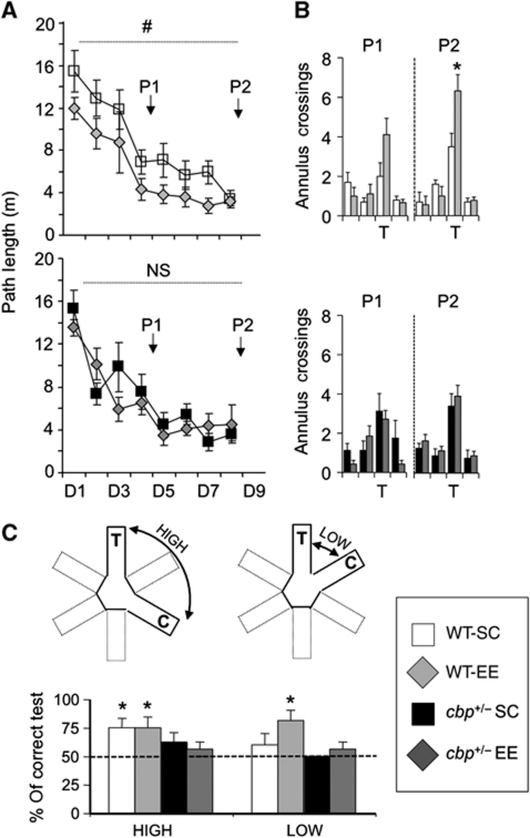
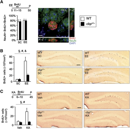
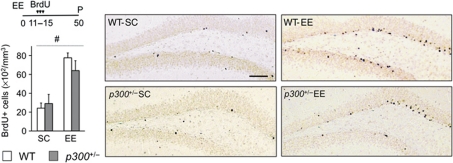
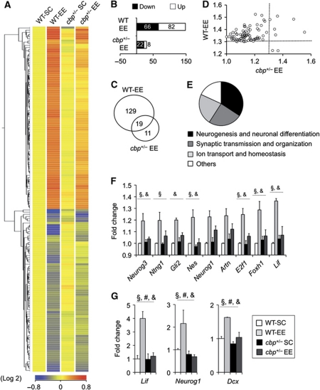
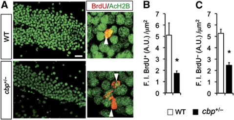
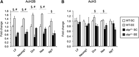
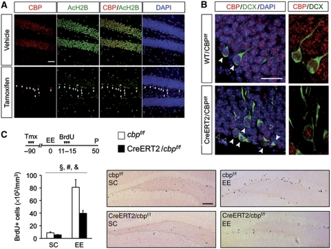
The epigenetic changes of the chromatin represent an attractive molecular substrate for adaptation to the environment. We examined here the role of CREB-binding protein (CBP), a histone acetyltransferase involved in mental retardation, in the genesis and maintenance of long-lasting systemic and behavioural adaptations to environmental enrichment (EE). Morphological and behavioural analyses demonstrated that EE ameliorates deficits associated to CBP deficiency. However, CBP-deficient mice also showed a strong defect in environment-induced neurogenesis and impaired EE-mediated enhancement of spatial navigation and pattern separation ability. These defects correlated with an attenuation of the transcriptional programme induced in response to EE and with deficits in histone acetylation at the promoters of EE-regulated, neurogenesis-related genes. Additional experiments in CBP restricted and inducible knockout mice indicated that environment-induced adult neurogenesis is extrinsically regulated by CBP function in mature granule cells. Overall, our experiments demonstrate that the environment alters gene expression by impinging on activities involved in modifying the epigenome and identify CBP-dependent transcriptional neuroadaptation as an important mediator of EE-induced benefits, a finding with important implications for mental retardation therapeutics.


Conflict of interest statement

The authors declare that they have no conflict of interest.

Figures

Figure 1
Figure 1
EE-mediated structural, behavioural and cognitive benefits. (A) Representative confocal images showing spines protruding from dendritic segments of hippocampal CA1 pyramidal neurons from cbp+/− mice and control littermates in SC or EE conditions. Scale bar: 5 μm. (B) Mice exposed to EE, independently of the genotype, show increased density of dendritic spines compared with animals kept in SC. Two-way ANOVA, #: significant housing effect; n=6–8 mice per group. These EE-dependent changes did not affect the morphology of dendritic spines (Supplementary Figure S1A–C) or the general cytoarchitecture of the hippocampus (Supplementary Figure S1D and E). (C) Cbp+/− mice show deficits in RotaRod performance and the exposure to EE causes a recovery in mutant mice and an improvement of the performance of WT mice. Two-way ANOVA, #: significant housing effect, §: significant genotype effect; n=10 mice per group. (D) Cbp+/− mice show an impairment in contextual fear conditioning. EE rescues this deficit and improves the contextual memory of WT mice. Two-way ANOVA, #: significant housing effect, §: significant genotype effect. t-Tests compared with WT-SC group: *P<0.05; **P<0.005; n=9–10 mice per group.
Figure 2
Figure 2
Cbp+/− mice show impaired EE-enhanced spatial navigation and pattern recognition ability. (A) The two-way ANOVA analysis of path lengths in the water maze task revealed a significant housing effect (F(1,31)housing=4.81, P=0.04), no genotype effect (F(1,31)genotype=0.37, P=0.55), and indicated a possible genotype × housing interaction (F(1,31)genotype × housing=3.53, P=0.07). More precisely, EE improved the performance in WT mice (upper panel, two-way ANOVA, #: significant housing effect), but not in cbp+/− mice (lower panel, NS: non-significant); n=8–10 mice per group. (B) EE housed wt mice showed more annulus crossings in the first and the second probe trials (upper panels: P1, P=0.06 no significant difference; P2, *P=0.03), whereas cbp+/− mice did not exhibit any housing effect (lower panels: P1, P=0.90; P2, P=0.70). (C) (Upper panels) Schematic representation of the WRM tests used to measure pattern separation. Mice were tested for their pattern separation ability by comparing their performance in two types of tests: low separation tests (LOW) in which the target arm (T, where the platform is located) and the choice arm (C) were contiguous, and high separation tests (HIGH) in which the target and the choice arms were separated by a closed arm. Graph: In the third day of training, the mice were subjected to two symmetrical low and high separation tasks and the average performance was calculated. WT mice housed in an enriched environment (WT-EE) performed well both kind of tests (percentage of correct HIGH: t(7)=2.65, P=0.03; percentage of correct LOW: t(7):3.42, P=0.01), whereas WT housed in standard cages (WT-SC) were only successful in the high separation tests (percentage of correct HIGH: WT-SC, t(9)=3.00, P=0.02). In contrast, cbp+/− mutants failed in both the HIGH and the LOW tests. *P<0.05 t-tests versus 50 (chance); n=8–10 mice per group.
Figure 3
Figure 3
Impaired induced neurogenesis in the SGZ of cbp+/− mice. (A) Animals received two daily injections of BrdU (100 mg/kg) for 5 consecutive days starting at day 11 of EE and were perfused (P) 35 days later. The right image shows cells in the SGZ immunolabelled with antibodies against BrdU (red) and NeuN (green) and nuclei counterstained with DAPI (blue). Five weeks after the last administration of BrdU, the vast majority of surviving cells were NeuN+. Scale bar: 2 μm. (B) CBP-deficient mice show a severe impairment in EE-induced neurogenesis in the SGZ. The right panels show representative images of BrdU (brown nuclei) immunostaining showing newborn neurons in the SGZ of WT and cbp+/− mice in SC and EE. Two-way ANOVA, #: significant housing effect, §: significant genotype effect, &: significant genotype × housing interaction; n=3–4 mice per group. Scale bars: 100 μm. (C) KA-induced neurogenesis is also impaired in cbp+/− mice. Six days after a single administration of KA (20 mg/kg) (KA) or vehicle (Veh), WT and cbp+/− mice received two daily BrdU injections for 5 consecutive days. Five weeks later, the mice were perfused (P) and adult newborn cells were stained for BrdU. The right panels show representative images of LRC immunolabelling in the SGZ of WT and cbp+/− mice treated with KA or vehicle. Two-way ANOVA, #: significant housing effect, §: significant genotype effect; n=4–5 per group. Scale bars: 100 μm.
Figure 4
Figure 4
Environment-induced neurogenesis is not altered in p300+/− mice. p300-deficient mice show normal basal and EE-induced neurogenesis in the SGZ. The right panels present representative images of BrdU (brown nuclei) immunostainings. Two-way ANOVA, #: significant housing effect; n=3–6 per group. Scale bar: 100 μm.
Figure 5
Figure 5
Impaired neuroadaptative transcriptional response to EE in the hippocampus of cbp+/− mice. (A) The hierarchical cluster of the 159 TCs differentially regulated in response to EE (corrected P-value <0.05, FC>1.3) reveals an attenuated transcriptional response in cbp+/− mice. (B) Number of TCs upregulated (white) and downregulated (black) in response to EE in WT and cbp+/− mice (referred to the respective SC groups) with FC>1.3. (C) Venn diagram showing the number of EE-regulated TCs. (D) Scatter plot comparing, in WT and cbp+/− mice, the FC of the 84 TCs differentially upregulated in response to EE. The dotted line indicates the threshold for FC. Most dots are located in the upper left quadrant, indicating that the changes are larger in WT animals. r(82)=0.31, P<0.05. (E) Pie diagram showing the number of unique entities associated to a GO term in each of the major functional categories identified in the analysis of gene sets differentially expressed in EE mice. (F) Bar graph showing the expression level of specific neurogenesis-related genes whose induction by EE is impaired in cbp+/− mice (expression values extracted from microarray data). Two-way ANOVA, §: significant genotype effect, &: significant genotype × housing interaction (non-corrected P-values). All these genes show a significant housing effect. Some interesting genes showing borderline significance are also presented. (G) qRT–PCR validation of EE-mediated hippocampal induction for the neurogenesis-related genes lif, neurog1 and dcx. Two-way ANOVA, #: significant housing effect, §: significant genotype effect, &: significant genotype × housing interaction; n=3 per group.
Figure 6
Figure 6
Reduced histone acetylation in the dentate gyrus of cbp+/− mice. (A) Representative confocal images of the DG labelled with BrdU (red) and acetylated histone H2B (green). Scale bar: 25 μm. (B) Quantification of fluorescence intensity in individual BrdU+ cells demonstrates that the acetylation of histone H2B is reduced in the proliferating neuroprogenitors of cbp+/− mice. *P<0.05 (unpaired two-tailed t-test), n=12–16 cells per group. (C) Quantification of fluorescence intensity in individual granule cells (BrdU cells adjacent to the BrdU+ cells shown in B) demonstrates a general reduction of histone H2B acetylation in the DG of cbp+/− mice. *P<0.05 (unpaired two-tailed t-test), n=46–50 cells per group.
Figure 7
Figure 7
Reduced histone acetylation at the promoters of neurogenesis-related genes. (A) ChIP assays using an antibody against AcH2B. (B) ChIP assays using an antibody against AcH3. Two-way ANOVA, #: significant housing effect, §: significant genotype effect; n=3 mice per sample, three samples per condition.
Figure 8
Figure 8
The environment-induced neurogenesis defect is not developmental and depends on intact levels of CBP in mature granule cells. (A) In forebrain-restricted inducible CBP knockout mice, tamoxifen injection causes the elimination of CBP immunoreactivity (red) in granule cells and severe hypoacetylation (green). The few remaining CBP+ cells in the inner blade of the DG show normal levels of AcH2B (arrow heads) and are NeuN (see Supplementary Figure S9), suggesting that only progenitors and newborn neurons still express CBP. Nuclei were stained with DAPI (blue). Scale bar: 50 μm. (B) Immunohistochemistry for CBP (red) and DCX (green) in coronal sections of CaMKIIa-creERT2/CBPf/f and WT/CBPf/f mice treated with tamoxifen demonstrate that most of the remaining CBP+ nuclei in the SGZ of CaMKIIa-creERT2/CBPf/f mice belong to Dcx+ cells (solid arrowheads). The empty arrowheads denote cells in the SGZ of the DG that were CBP+ and Dcx. Nuclei were stained with DAPI (blue). Right, magnification of the dotted squares in the left images show CBP+ and DCX+ cells in the SGZ of the DG. Scale bar: 15 μm. (C) Tamoxifen was administered to 2-month-old mice and 12 weeks later, mice were housed in EE or maintained in SC. Animals received two daily injections of BrdU (100 mg/kg) for 5 consecutive days starting at day 11 of EE. Five weeks after the last administration of BrdU, the animals were perfused (P) for immunohistochemistry. CaMKIIa-creERT2/CBPf/f mice show a severe impairment in EE-induced neurogenesis in the SGZ. The right panels show representative images of BrdU (brown nuclei) immunostaining showing newborn neurons in the SGZ of CaMKIIa-creERT2/CBPf/f mice and control littermates housed in either SC or EE. Two-way ANOVA, #: significant housing effect, §: significant genotype effect, &: significant genotype × housing interaction; n=4–5 per group. Scale bar: 100 μm.

References

    1. Aimone JB, Deng W, Gage FH (2011) Resolving new memories: a critical look at the dentate gyrus, adult neurogenesis, and pattern separation. Neuron 70: 589–596 - PMC - PubMed
    1. Alarcon JM, Malleret G, Touzani K, Vronskaya S, Ishii S, Kandel ER, Barco A (2004) Chromatin acetylation, memory, and LTP are impaired in CBP+/− mice: a model for the cognitive deficit in Rubinstein-Taybi syndrome and its amelioration. Neuron 42: 947–959 - PubMed
    1. Arai JA, Li S, Hartley DM, Feig LA (2009) Transgenerational rescue of a genetic defect in long-term potentiation and memory formation by juvenile enrichment. J Neurosci 29: 1496–1502 - PMC - PubMed
    1. Barco A (2007) The Rubinstein-Taybi syndrome: modeling mental impairment in the mouse. Genes Brain Behav 6: 32–39 - PubMed
    1. Barrett RM, Malvaez M, Kramar E, Matheos DP, Arrizon A, Cabrera SM, Lynch G, Greene RW, Wood MA (2011) Hippocampal focal knockout of CBP affects specific histone modifications, long-term potentiation, and long-term memory. Neuropsychopharmacology 36: 1545–1556 - PMC - PubMed

Publication types