. 2011 Aug 16:7:521.
doi: 10.1038/msb.2011.55.

Engineering microbes to sense and eradicate Pseudomonas aeruginosa, a human pathogen

Affiliations

Engineering microbes to sense and eradicate Pseudomonas aeruginosa, a human pathogen

Nazanin Saeidi et al. Mol Syst Biol. .

Abstract

Synthetic biology aims to systematically design and construct novel biological systems that address energy, environment, and health issues. Herein, we describe the development of a synthetic genetic system, which comprises quorum sensing, killing, and lysing devices, that enables Escherichia coli to sense and kill a pathogenic Pseudomonas aeruginosa strain through the production and release of pyocin. The sensing, killing, and lysing devices were characterized to elucidate their detection, antimicrobial and pyocin release functionalities, which subsequently aided in the construction of the final system and the verification of its designed behavior. We demonstrated that our engineered E. coli sensed and killed planktonic P. aeruginosa, evidenced by 99% reduction in the viable cells. Moreover, we showed that our engineered E. coli inhibited the formation of P. aeruginosa biofilm by close to 90%, leading to much sparser and thinner biofilm matrices. These results suggest that E. coli carrying our synthetic genetic system may provide a novel synthetic biology-driven antimicrobial strategy that could potentially be applied to fighting P. aeruginosa and other infectious pathogens.


Conflict of interest statement

The authors declare that they have no conflict of interest.

Figures

Figure 1
Figure 1
Schematic of ‘Pathogen Sensing and Killing’ system. luxR promoter is activated by LasR-3OC12HSL complex, leading to production of E7 lysis protein and S5 pyocin within E. coli chassis. After the E7 protein attains the threshold concentration that causes the chassis to lyse, the accumulated S5 is released into the exogenous environment and kills P. aeruginosa.
Figure 2
Figure 2
Characterization results of sensing device coupled with GFP reporter. (A) GFP production rate per cell over time at different 3OC12HSL inducer concentrations. (B) Time-averaged GFP production rate per cell at different input 3OC12HSL concentrations, showing that the optimal operating concentrations for the sensing device range from 1.0E-7 to 1.0E-6 M 3OC12HSL. Error bar represents the standard deviation of statistical means between 20 and 80 m after induction. The experiment was performed with six replicates. Source data is available for this figure at www.nature.com/msb.
Figure 3
Figure 3
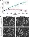
Characterization results of lysis device using 3OC12HSL. (A) Growth curve of E. coli expressing E7 lysis protein after induction with different concentrations of 3OC12HSL. (B, C) Effects of lysis protein on E. coli surface morphology as observed using FESEM. The results show that the surface of the E. coli was damaged when E. coli carrying pTetR-LasR-pLuxR-E7 and E. coli carrying pTetR-LasR-pLuxR-S5-pLuxR-E7 (the final system) were induced with 3OC12HSL. Scale bar: 1 μm. Error bar represents the standard deviation of four replicates. Source data is available for this figure at www.nature.com/msb.
Figure 4
Figure 4
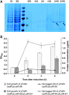
Characterization results of the lysis device in the final system using 3OC12HSL. (A) SDS–PAGE of (i, ii) total extracellular proteins and (iii–viii) IMAC purified His-tagged S5 protein sampled from the extracellular supernatant. Total extracellular proteins exported from (i) E. coli carrying pTetR-LasR-pLuxR-S5 (without lysis device) was significantly lesser than that exported from (ii) E. coli carrying pTetR-LasR-pLuxR-S5-pLuxR-E7 (the final system) as indicated in darker lanes of (ii) relative to (i). (iii–v): E. coli carrying pTetR-LasR-pLuxR-S5 (without lysis device) at 0, 2, and 4 h after induction. (vi–viii) E. coli carrying pTetR-LasR-pLuxR-S5-pLuxR-E7 (the final system) at 0, 2, and 4 h after induction. The results show that pyocin S5 (57 kDa; arrowed) was only detectable in lanes that corresponded to E. coli carrying the final system and not in lanes of E. coli without the lysis device. Ladder used was Bio-Rad's Precision Plus Protein standards. (B) Characterization of lysis device in the final system by optical density (bar graphs) and concentration of pyocin released (lines) after induction. The results show an impulse release of pyocin S5 at 2 h after induction, followed by a sustained steady-state release in the final system (dotted lines). Optical density of the final system was characterized by an initial decrease at 2 h after induction, indicative of the onset of lysis, after which the regrowth of engineered E. coli occurs (shaded bar). Correspondingly, the concentration of pyocin released in E. coli without the lysis device (solid line) was 1/8 that of the final system with a continually increasing optical density (unshaded bar). Error bar represents the standard deviation of two replicates. Source data is available for this figure at www.nature.com/msb.
Figure 5
Figure 5
Inhibition of P. aeruginosa by the engineered E. coli induced with native 3OC12HSL produced by P. aeruginosa. (A) Agar overlay assay of P. aeruginosa after exposure to supernatant of four different cultures. First, P. aeruginosa exposed to supernatant of wild-type E. coli showed no bactericidal activity. Second, P. aeruginosa exposed to supernatant of wild-type E. coli mixed with P. aeruginosa produced no inhibition zones. Third, exposure to supernatant of E. coli carrying pTetR-LasR-pLuxR-S5-pLuxR-E7 (final system) did not produce any inhibition as well. Fourth, only P. aeruginosa exposed to supernatant of E. coli carrying final system with P. aeruginosa displayed clear inhibition zones, which suggested that the system produced sufficient pyocin S5 to exhibit bactericidal activity. (B) P. aeruginosa cells stained using the LIVE/DEAD cell viability assay. Many P. aeruginosa cells were stained with PI dye, which indicate dead cells, when exposed to supernatant of engineered E. coli carrying the final system that was induced by native 3OC12HSL produced by P. aeruginosa. Scale bar: 5 μm. (C) Fluorescence measurement of P. aeruginosa that constitutively expresses GFP in mixed culture with engineered E. coli. The result from the mixed culture with the engineered E. coli carrying pTetR-LasR-pLuxR-E7 and pTet-LasR-pLuxR-S5 shows an exponential increase in the fluorescence readings, whereas the mixed culture with E. coli carrying pTetR-LasR-pLuxR-S5-pLuxR-E7 (the final system) exhibited no increase in the readings. This suggests that the growth of P. aeruginosa was significantly inhibited in the mixed culture with engineered E. coli carrying the final system. PAO1, which pyocin S5 was derived from, was included as a negative control. Error bar represents the standard deviation of six replicates. (D) Percentage survival of P. aeruginosa carrying chloramphenicol-resistant plasmid in mixed culture with the engineered E. coli. Pseudomonas in the mixed culture was quantified by viable cell count using chloramphenicol selection. The result shows that our engineered E. coli inhibited the growth of Pseudomonas by 99%. In contrast, inhibition was less observed in Pseudomonas co-cultured with incomplete E. coli systems missing either the pyocin S5 killing device or E7 lysis device. Error bar represents the standard deviation of three replicates. Source data is available for this figure at www.nature.com/msb.
Figure 6
Figure 6
Biofilm inhibition assay with engineered E. coli. (A) Percentage survival of P. aeruginosa biofilm carrying chloramphenicol-resistant plasmid. Pseudomonas biofilm was grown in a polystyrene 24-well plate in the presence of the engineered E. coli for 18 h and quantified by viable cell count using chloramphenicol selection. The results imply that the formation of Pseudomonas biofilm was inhibited by close to 90% with the engineered E. coli carrying the final system (pTetR-LasR-pLuxR-S5-pLuxR-E7) as compared with biofilm grown with wild-type E. coli or incomplete E. coli system missing either pyocin S5 or E7 lysis genes. P. aeruginosa PAO1, which pyocin S5 was derived from, was included as a negative control. Error bar represents the standard deviation of six replicates. (B) Biofilm inhibition observed under CLSM microscopy. Pseudomonas biofilm with green fluorescence was grown on glass slide in the presence of the engineered E. coli and visualized under CLSM microscope after 18 h. Images reconstructed from biofilm Z-stacks using Zeiss 2.5D software implied that the initialization and progression of biofilm cells into multilayers were strongly inhibited for Pseudomonas grown with E. coli carrying the final system as opposed to lush and elaborated biofilm formation observed in Pseudomonas grown alone or with incomplete E. coli system missing either pyocin S5 or E7 lysis genes. Scale bar: 50 μm. Z-stack: 40 μm. Source data is available for this figure at www.nature.com/msb.

Comment in

References

    1. Anderson JC, Clarke EJ, Arkin AP, Voigt CA (2006) Environmentally controlled invasion of cancer cells by engineered bacteria. J Mol Biol 355: 619–627 - PubMed
    1. Asad S, Opal SM (2008) Bench-to-bedside review: quorum sensing and the role of cell-to-cell communication during invasive bacterial infection. Crit Care 12: 236. - PMC - PubMed
    1. Baba T, Schneewind O (1998) Instruments of microbial warfare: bacteriocin synthesis, toxicity and immunity. Trends Microbiol 6: 66–71 - PubMed
    1. Bradford MM (1976) A rapid and sensitive method for the quantitation of microgram quantities of protein utilizing the principle of protein-dye binding. Anal Biochem 72: 248–254 - PubMed
    1. Brook I (2005) The role of bacterial interference in otitis, sinusitis and tonsillitis. Otolaryngol Head Neck Surg 133: 139–146 - PubMed

Publication types

MeSH terms