Case Reports
. 2011:5:1083-8.
doi: 10.2147/OPTH.S22786. Epub 2011 Aug 4.

Central retinal artery occlusion resembling Purtscher-like retinopathy

Affiliations
Case Reports

Central retinal artery occlusion resembling Purtscher-like retinopathy

Takuji Kurimoto et al. Clin Ophthalmol. 2011.

Abstract

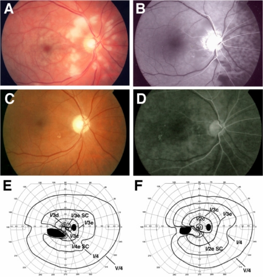
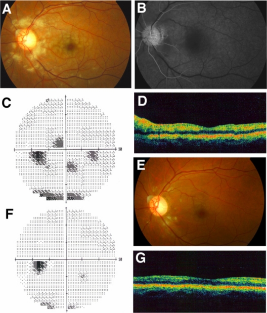
This paper reports three cases of central retinal artery occlusion (CRAO) with Purtscher-like retinopathy and good recovery of visual function. The three cases of CRAO had similar fundus changes, ie, cotton wool patches surrounding the optic disc and whitening of the retina surrounding the fovea with a cherry red spot. Fluorescein angiography showed a delay of arm-to-retina circulation time and a partial defect of choroid circulation. Although the three cases were treated by different regimens of steroid pulse therapy and antiplatelet therapy, visual function recovered well and all disturbances of the retinal and choroid circulations resolved. Although eyes with a CRAO normally have a poor visual prognosis, our three cases responded well to the treatments and recovered good visual function. Thus, cases showing fundus changes similar to our three cases may have a pathogenesis different from that of a complete CRAO.

Keywords: Purtscher retinopathy; central retinal artery occlusion; cotton wool patches; steroid therapy.


Figures

Figure 1
Figure 1
Clinical findings for Case 1. Fundus photographs and fluorescent angiograms before treatment. There are many cotton wool patches surrounding the optic disc and retinal whitening surrounding the fovea (A). Fluorescein angiography shows nonperfused areas of retinal arterioles in the areas corresponding to the areas of cotton wool patches. A delay in the choroidal filling in the nasal area and arm-to-retina time (32 seconds) was found (B). Fundus photograph (C) and fluorescent angiogram (D) after treatment. Optic disc is slightly pale, and defects of the nerve fiber layer can be seen (C). Fluorescein angiography shows improvement of the retinal circulation (D). Arm-to-retina time was 15 seconds. Goldmann kinetic perimetry before (E) and after (F) treatment. There is a large absolute scotoma in the lower nasal field and paracentral scotoma before treatment (E). The paracentral scotoma is decreased after treatment (F). Note: Black indicates absolute scotoma and gray relative scotoma in E and F. Abbreviation: SC, scotoma.
Figure 2
Figure 2
Clinical findings for Case 2. Fundus photographs and fluorescein angiograms before treatment. There are many cotton wool patches surrounding the optic disc and retinal whitening surrounding the fovea (A). Fluorescein angiography showed nonperfused areas of retinal arterioles in areas corresponding to the cotton wool patches. The arm-to-retina time was 28 seconds (B). Humphrey static perimetry at onset (C) and 1 month later (F). Paracentral scotomas are almost gone. Fundus photograph 1 month after the onset (E). Most of the cotton wool patches and retinal whitening have disappeared. Optical coherence tomography at onset (D) and 1 month later (G). Swelling of the inner retina showing hyper-reflectivity was found at onset (D), but the hyper-reflectivity has disappeared and retinal thickness returned to normal 1 month after onset (G).
Figure 3
Figure 3
Clinical findings for Case 3. Fundus photographs and fluorescein angiograms before treatment. There are many cotton wool patches surrounding the optic disc and slight retinal whitening surrounding the fovea (A). Fluorescein angiography showing that nonperfused areas of the retinal arterioles were found in the areas corresponding to the cotton wool patches. The delay in choroidal filling in the nasal area and arm-to-retina time (28 seconds) were found (B). Goldmann kinetic perimetry before (C) and 6 weeks after (D) treatment. There is a central scotoma and a constriction in lower nasal field and paracentral scotoma before treatment (C). Central scotoma is decreased and the constriction of lower nasal field has abated after treatment (D). Fundus photograph (C) and fluorescein angiogram (D) 6 weeks after treatment. Cotton wool patches are not present and optic disc is slightly pale (E). Fluorescein angiography showed improvement of retinal circulation 38 seconds after the injection of fluorescein (D). Note: Black indicates absolute scotoma and gray relative scotoma in C and D. Abbreviation: SC, scotoma.

References

    1. Hayreh SS, Zimmerman MB. Fundus changes in central retinal artery occlusion. Retina. 2007;27(3):276–289. - PubMed
    1. Augsburger JJ, Magargal LE. Visual prognosis following treatment of acute central retinal artery obstruction. Br J Ophthalmol. 1980;64(12):913–917. - PMC - PubMed
    1. Brown GC, Shields JA. Cilioretinal arteries and retinal arterial occlusion. Arch Ophthalmol. 1979;97(1):84–92. - PubMed
    1. Brown GC, Magargal LE. Central retinal artery obstruction and visual acuity. Ophthalmology. 1982;89(1):14–19. - PubMed
    1. Yuzurihara D, Iijima H. Visual outcome in central retinal and branch retinal artery occlusion. Jpn J Ophthalmol. 2004;48(5):490–492. - PubMed

Publication types