

Detection of colorectal adenomas using a bioactivatable probe specific for matrix metalloproteinase activity
- PMID: 21847360
- PMCID: PMC3156659
- DOI: 10.1593/neo.11400
Detection of colorectal adenomas using a bioactivatable probe specific for matrix metalloproteinase activity
Abstract
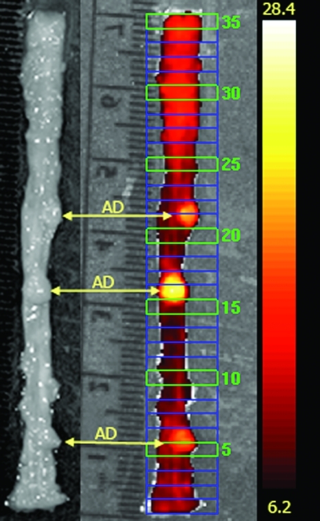
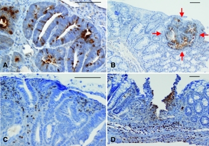
A significant proportion of colorectal adenomas, in particular those that lack an elevated growth component, continue to escape detection during endoscopic surveillance. Elevation of the activity of matrix metalloproteinases (MMPs), a large family of zinc endopeptidases, in adenomas serves as a biomarker of early tumorigenesis. The goal of this study was to assess the feasibility of using a newly developed near-infrared bioactivatable probe (MMPSense 680) that reports the activity of a broad array of MMP isoforms to detect early colorectal adenomas. Adenomatous polyposis coli (Apc)(+/Min-FCCC) mice that spontaneously develop multiple colorectal adenomas were injected with MMPSense 680, and the colons were imaged in an IVIS Spectrum system ex vivo. Image analyses were correlated with histopathologic findings for all regions of interest (ROIs). The biochemical basis of fluorescent signal was investigated by immunohistochemical staining of MMP-7 and -9. A strong correlation (Kendall = 0.80) was observed between a positive signal and the presence of pathologically confirmed colonic adenomas; 92.9% of the 350 ROIs evaluated were classified correctly. The correlation between two independent observers was 0.87. MMP-7 expression was localized to epithelial cells of adenomas and microadenomas, whereas staining of MMP-9 was found in infiltrating polymorphonuclear leukocytes within the adenomas. MMPSense 680 identifies colorectal adenomas, both polypoid and nonpolypoid, in Apc(+/Min-FCCC) mice with high specificity. Use of this fluorescent probe in combination with colonoscopy could aid in preventing colorectal neoplasias by providing new opportunities for early detection and therapeutic intervention.
Figures
Similar articles
-
Immunohistochemical expression of matrix metalloproteinase-7 in human colorectal adenomas using specified automated cellular image analysis system: a clinicopathological study.Saudi J Gastroenterol. 2013 Jan-Feb;19(1):23-7. doi: 10.4103/1319-3767.105916. Saudi J Gastroenterol. 2013. PMID: 23319034 Free PMC article.
-
Matrix metalloproteinase-13 expression in the progression of colorectal adenoma to carcinoma : Matrix metalloproteinase-13 expression in the colorectal adenoma and carcinoma.Tumour Biol. 2014 Jun;35(6):5653-8. doi: 10.1007/s13277-014-1748-9. Epub 2014 Feb 23. Tumour Biol. 2014. PMID: 24563279
-
Active matrix metalloproteinase-2 activity discriminates colonic mucosa, adenomas with and without high-grade dysplasia, and cancers.Hum Pathol. 2011 May;42(5):688-701. doi: 10.1016/j.humpath.2010.08.021. Epub 2011 Jan 15. Hum Pathol. 2011. PMID: 21237495 Free PMC article.
-
Novel clinical in vivo roles for indigo carmine: high-magnification chromoscopic colonoscopy.Biotech Histochem. 2007 Apr;82(2):57-71. doi: 10.1080/10520290701259340. Biotech Histochem. 2007. PMID: 17577700 Review.
-
The unique pathology of nonpolypoid colorectal neoplasia in IBD.Gastrointest Endosc Clin N Am. 2014 Jul;24(3):455-68. doi: 10.1016/j.giec.2014.03.009. Gastrointest Endosc Clin N Am. 2014. PMID: 24975536 Review.
Cited by
-
Impact of proteolytic enzymes in colorectal cancer development and progression.World J Gastroenterol. 2014 Oct 7;20(37):13246-57. doi: 10.3748/wjg.v20.i37.13246. World J Gastroenterol. 2014. PMID: 25309062 Free PMC article. Review.
-
Up-regulation of microRNA-302a inhibited the proliferation and invasion of colorectal cancer cells by regulation of the MAPK and PI3K/Akt signaling pathways.Int J Clin Exp Pathol. 2015 May 1;8(5):4481-91. eCollection 2015. Int J Clin Exp Pathol. 2015. PMID: 26191138 Free PMC article.
-
Chemical biology for understanding matrix metalloproteinase function.Chembiochem. 2012 Sep 24;13(14):2002-20. doi: 10.1002/cbic.201200298. Epub 2012 Aug 30. Chembiochem. 2012. PMID: 22933318 Free PMC article. Review.
-
Phthalocyanine-Blue Nanoparticles for the Direct Visualization of Tumors with White Light Illumination.ACS Appl Mater Interfaces. 2023 Jul 19;15(28):33373-33381. doi: 10.1021/acsami.3c05140. Epub 2023 Jul 3. ACS Appl Mater Interfaces. 2023. PMID: 37395349 Free PMC article.
-
In vivo investigation of hybrid Paclitaxel nanocrystals with dual fluorescent probes for cancer theranostics.Pharm Res. 2014 Jun;31(6):1450-9. doi: 10.1007/s11095-013-1048-x. Epub 2013 Apr 26. Pharm Res. 2014. PMID: 23619595
References
-
- Centers for Disease Control and Prevention, author. Vital signs: colorectal cancer screening among adults aged 50–75 years — United States, 2008. MMWR Morb Mortal Wkly Rep. 2010;59:808–812. - PubMed
-
- Heresbach D, Barrioz T, Lapalus MG, Coumaros D, Bauret P, Potier P, Sautereau D, Boustiere C, Grimaud JC, Barthelemy C, et al. Miss rate for colorectal neoplastic polyps: a prospective multicenter study of back-to-back video colonoscopies. Endoscopy. 2008;40:284–290. - PubMed
-
- Cornett D, Barancin C, Roeder B, Reichelderfer M, Frick T, Gopal D, Kim D, Pickhardt PJ, Taylor A, Pfau P. Findings on optical colonoscopy after positive CT colonography exam. Am J Gastroenterol. 2008;103:2068–2074. - PubMed
-
- Soetikno RM, Kaltenbach T, Rouse RV, Park W, Maheshwari A, Sato T, Matsui S, Friedland S. Prevalence of nonpolypoid (flat and depressed) colorectal neoplasms in asymptomatic and symptomatic adults. JAMA. 2008;299:1027–1035. - PubMed
Publication types
MeSH terms
Substances
Grants and funding
LinkOut - more resources
Full Text Sources
Other Literature Sources
Medical
Miscellaneous