. 2011 Aug;13(8):758-70.
doi: 10.1593/neo.11528.

Type III TGF-β receptor enhances colon cancer cell migration and anchorage-independent growth

Affiliations

Type III TGF-β receptor enhances colon cancer cell migration and anchorage-independent growth

Catherine E Gatza et al. Neoplasia. 2011 Aug.

Abstract

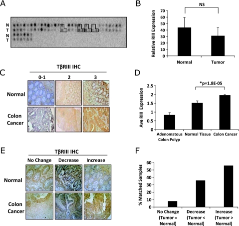
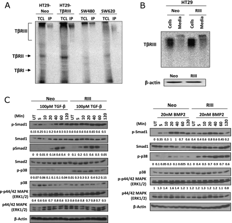
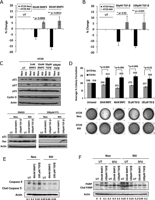
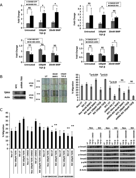
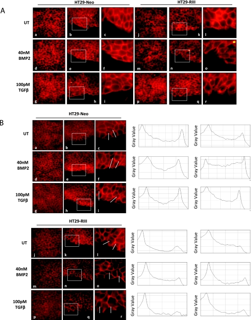
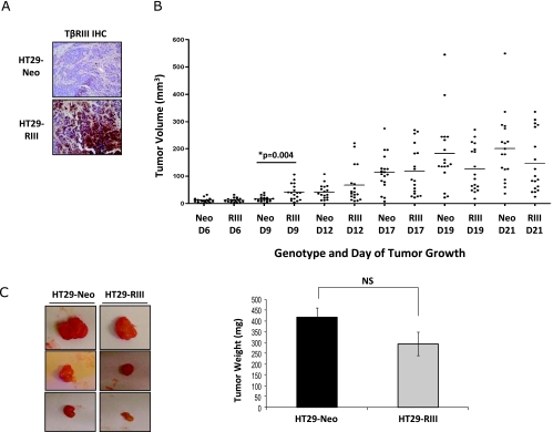
The type III TGF-β receptor (TβRIII or betagylcan) is a TGF-β superfamily coreceptor with emerging roles in regulating TGF-β superfamily signaling and cancer progression. Alterations in TGF-β superfamily signaling are common in colon cancer; however, the role of TβRIII has not been examined. Although TβRIII expression is frequently lost at the message and protein level in human cancers and suppresses cancer progression in these contexts, here we demonstrate that, in colon cancer, TβRIII messenger RNA expression is not significantly altered and TβRIII expression is more frequently increased at the protein level, suggesting a distinct role for TβRIII in colon cancer. Increasing TβRIII expression in colon cancer model systems enhanced ligand-mediated phosphorylation of p38 and the Smad proteins, while switching TGF-β and BMP-2 from inhibitors to stimulators of colon cancer cell proliferation, inhibiting ligand-induced p21 and p27 expression. In addition, increasing TβRIII expression increased ligand-stimulated anchorage-independent growth, a resistance to ligand- and chemotherapy-induced apoptosis, cell migration and modestly increased tumorigenicity in vivo. In a reciprocal manner, silencing endogenous TβRIII expression decreased colon cancer cell migration. These data support a model whereby TβRIII mediates TGF-β superfamily ligand-induced colon cancer progression and support a context-dependent role for TβRIII in regulating cancer progression.


Figures

Figure 1
Figure 1
TβRIII expression increases during colon cancer. (A) A cDNA array with matched normal and colon tumors (n = 37) was hybridized with a [32P]-labeled probe for TβRIII. The signal intensity for each spot was determined using ImageJ software. Boxed areas represent paired normal, tumor, and metastases. (B) Graphical representation of the mean signal intensity ± SD of the intensity. NS indicates not significant. (C) TβRIII IHC was performed on a human tissue microarray of normal colon and tumor specimens. The tissue array was scored on a scale of 0 (no staining) to 3 (highest). Adenomatous colon polyp (n = 34), normal tissue (n = 60), and colon cancer (n = 323). Original magnification, x20. (D) Graphical representation of the average intensity score of TβRIII protein expression in normal and tumor tissues ± SEM. (E) TβRIII IHC was performed on a human tissue microarray of normal colon and tumor specimens. Representative matched normal and tumor sample pairs from the same patient are shown, demonstrating no change, an increase, or a decrease in TβRIII expression. Original magnification, x20. (F) Graphical representation of the percentage of matched pairs (n = 25) that demonstrate an increase (n = 14), no change (n = 2), or decrease (n = 9) in TβRIII protein expression in tumor versus normal tissue.
Figure 2
Figure 2
TβRIII is expressed in colon cancer cell lines and enhances BMP-2- and TGF-β-mediated signaling. (A) Binding and cross-linking of total cell lysates (TCL) (left lanes) and immunoprecipitation (IP) for TβRIII (right lanes) from four different cell lines: HT29 stable cell lines, namely, HT29-Neo and HT29-TβRIII; SW480 cells, derived from a primary colorectal adenocarcinoma; and SW620 cells, derived from the metastatic lymph node site of a colorectal adenocarcinoma from the same patient as the SW480 line. (B) Expression of sTβRIII in HT29 stable cell lines. Cells were grown in 10% FBS McCoy 5A for 24 hours. TβRIII and sTβRIII expression was examined in cells and medium by binding and cross-linking. β-Actin serves as a total protein control (bottom panel). (C) HT29-Neo and TβRIII cells were serum-starved overnight and then treated with 100 pM TGF-β or 20 nM BMP-2 during the indicated time course (minutes). Western blot analyses were performed for the indicated proteins. Densitometric analysis is shown normalized to β-actin.
Figure 3
Figure 3
TβRIII expression increases colon cancer in vitro tumorigenicity. (A) HT29-Neo and TβRIII cells were treated with 20 or 40 nM BMP-2 for 24 hours. Proliferation was analyzed by a 3H incorporation assay. The percent proliferation was determined by normalizing the counts to those of untreated samples. (B) HT29-Neo and TβRIII cells were treated with 50 or 100 pM TGF-β1 for 24 hours. Proliferation was analyzed by a 3H incorporation assay. (C) HT29-Neo and TβRIII cells were treated with 2 or 10 nM BMP-2 and 50 or 100 pM TGF-β1 for 24 hours. HT29-Neo and TβRIII cells were treated with 200 µM FTS or DMSO for 3 days. Western blot analyses were performed to analyze protein levels of p21, p27, p15, cyclin D, and Ras with β-actin as a total protein control. (D) HT29-Neo and TβRIII cells were plated in a soft agar assay untreated or treated with 20 nM BMP-2, 40 nM BMP-2, 50 pM TGF-β, or 100 pM TGF-β for 21 days. The mean percent colony formation ± SEM is shown normalized to the untreated Neo or TβRIII. Average colony number is shown above the bar graph. (E) HT29-Neo and TβRIII cells were treated with 40 nM BMP-2 or 100 pM TGF-β for 48 hours and examined for apoptosis by Western blot analysis of caspase 9 levels. Densitometric analysis is shown normalized to β-actin. (F) HT29-Neo and TβRIII cells were treated with 50 µM 5-fluorouracil for 48 hours. Cells were concurrently treated with 20 nM BMP-2 or 100 pM TGF-β and examined for apoptosis induction by Western blot analysis of PARP cleavage. Densitometric analysis is shown normalized to β-actin.
Figure 4
Figure 4
TβRIII increases colon cancer cell migration. (A) SW480 and SW620 GFP, TβRIII, NTC, or shTβRIII adenovirally infected cells were plated in a fibronectin transwell migration assay. Cells were plated in serum-free conditions on a fibronectin-coated transwell (50 µg/ml) with and without ligand treatment. Migration toward serum was measured by counting the number of cells on the filter after 12 hours. Fold change ± SEM is demonstrated. * P < .05. NS indicates not significant. (B) HT29-Neo, TβRIII, and NTC (nontargeting control) or shRNA TβRIII adenovirally infected cells were plated in a monolayer scratch wound assay. Cells were grown to confluence and then wounded by scratching and treated with 40 nM BMP-2 or 100 pM TGF-β. The percent migration was calculated by measuring the wound closure over time (0 and 24 hours). (C) Scratch wound assay with HT29-Neo and TβRIII cells treated with ligand and the ALK5 inhibitor SB431542 (5 µM) or the p38 inhibitor SB203580 (15 µM). The percent migration was calculated by measuring the wound closure over time (0 and 24 hours). *P = .02 RIII UT versus RIII UT+SB431542. **P=.01 RIII 100 pMTGF-β versus RIII 100 pM TGF-β + SB431542, RIII UT versus RIII UT + SB203580, RIII 100 pM TGF-β versus RIII 100 pM TGF-β + SB203580. Western blot analysis showing inhibition of TGF-β signaling with inhibitor treatment. Cells were treated with 100 pM TGF-β for 40 minutes, with or without DMSO, 5 µM SB431542 or 15 µM SB203580 treatment.
Figure 5
Figure 5
TβRIII alters actin and E-cadherin localization in colon cancer cells. (A) Actin (phalloidin) immunofluorescent staining. HT29-Neo and TβRIII cells were grown to confluence and then wounded by scratching and treated with 40 nM BMP-2 or 100 pM TGF-β. At 18 hours after scratch, cells were fixed and stained for actin. Images show actin staining in confluent culture (a, d, g, j, m, p) and along the wound edge (b, e, h, k, n, q). Original magnification, x60. Boxed area (b, e, h, k, n, q) is shown enlarged in c, f, i, l, o, r. (B) E-cadherin immunofluorescent staining. HT29-Neo and TβRIII cells were grown to confluence and then wounded by scratching and treated with 40 nM BMP-2 or 100 pM TGF-β. At 18 hours after scratch, cells were fixed and stained for E-cadherin. Images show E-cadherin staining in confluent culture (a, d, g, j, m, p) and along the wound edge (b, e, h, k, n, q). Original magnification, x60. Boxed area (b, e, h, k, n, q) is shown enlarged in c, f, i, l, o, r. Line scan analysis of individual cells (ImageJ software; NIH) shows E-cadherin staining intensity in a single cell from the interior toward the wound edge. The line demonstrates the area of measured intensity.
Figure 6
Figure 6
TβRIII enhances early colon cancer tumorigenicity in vivo. A total of 1 x 10⁶ HT29-Neo or HT29-TβRIII colon cancer cells were injected subcutaneously into the right and left flanks of BALB/cAnNCr nu/nu mice. Mice were weighed, and tumor width (W) and length (L) were measured every 3 days. Tumor volume was determined using the formula: V = 0.5 x L x W2. (A) TβRIII IHC of HT29-Neo and TβRIII tumors at day 21. (B) Graphical representation of HT29-Neo and TβRIII tumor volume over time (D indicates day). D9, *P = .004. (C) Representative images of HT29-Neo and TβRIII xenograft tumors and graphical comparison of final tumor mass ± SEM of HT29-Neo and TβRIII xenografts at day 21. NS indicates not significant.

References

    1. Massague J. TGFβ in cancer. Cell. 2008;34:215–230. - PubMed
    1. Pardali K, Moustakas A. Actions of TGF-β as tumor suppressor and pro-metastatic factor in human cancer. Biochim Biophys Acta. 2007;1775:21–62. - PubMed
    1. Esparza-Lopez J, Montiel JL, Vilchis-Landeros MM, Okadome T, Miyazono K, Lopez-Casillas F. Ligand binding and functional properties of betaglycan, a co-receptor of the transforming growth factor-β superfamily. Specialized binding regions for transforming growth factor-β and inhibin A. J Biol Chem. 2001;276:14588–14596. - PubMed
    1. Gatza CE, Oh SY, Blobe GC. Roles for the type III TGF-β receptor in human cancer. Cell Signal. 2010;22:1163–1174. - PMC - PubMed
    1. Kirkbride KC, Townsend TA, Bruinsma MW, Barnett JV, Blobe GC. Bone morphogenetic proteins signal through the transforming growth factor-β type III receptor. J Biol Chem. 2008;283:7628–7637. - PubMed

Publication types

MeSH terms

Substances

LinkOut - more resources