Comparative Study
. 2011 Nov;54(11):2931-40.
doi: 10.1007/s00125-011-2272-8. Epub 2011 Aug 17.

LDL from obese patients with the metabolic syndrome show increased lipid peroxidation and activate platelets

Affiliations
Comparative Study

LDL from obese patients with the metabolic syndrome show increased lipid peroxidation and activate platelets

R Colas et al. Diabetologia. 2011 Nov.

Abstract

Aims/hypothesis: This study assessed oxidative stress in LDL from obese patients with the metabolic syndrome and compared it with that in LDL from type 2 diabetic patients or control volunteers. It also determined the effect on platelets of LDL from the three groups.

Methods: The profiles of lipids, fatty acids and fatty acid oxidation products were determined in LDL isolated from plasma of patients with the metabolic syndrome, patients with type 2 diabetes and volunteers (n = 10 per group). The effects of LDL from the participant groups on the platelet arachidonic acid signalling cascade and aggregation were investigated.

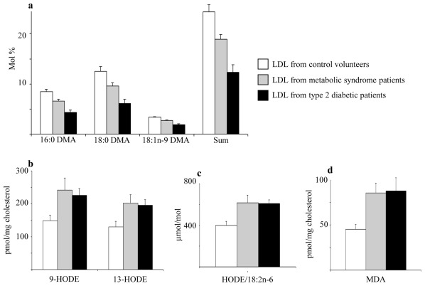
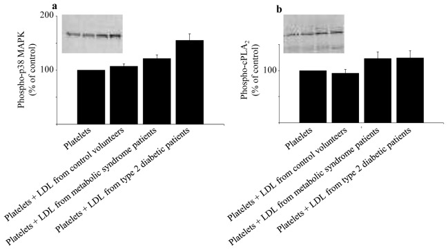
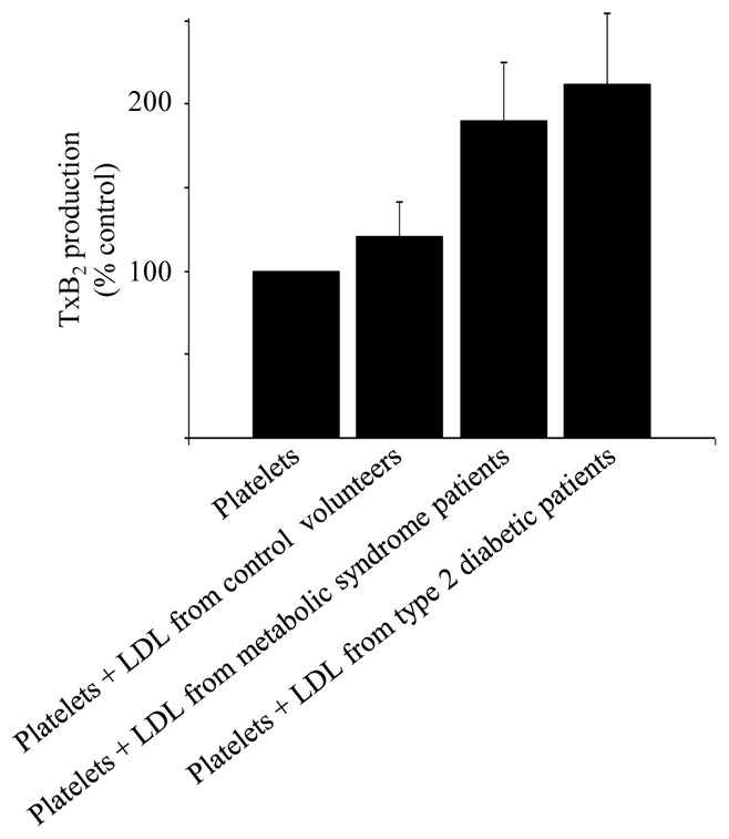
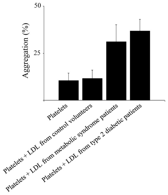
Results: Compared with LDL from control volunteers, LDL from obese metabolic syndrome and type 2 diabetic patients had lower cholesteryl ester, higher triacylglycerol and lower ethanolamine plasmalogen levels. Proportions of linoleic acid were decreased in phosphatidylcholine and cholesteryl esters in LDL from both patient groups. Among the markers of lipid peroxidation, oxidation products of linoleic acid (hydroxy-octadecadienoic acids) and malondialdehyde were increased by 59% and twofold, respectively in LDL from metabolic syndrome and type 2 diabetic patients. LDL from metabolic syndrome and type 2 diabetic patients were equally potent in activating the platelet arachidonic acid signalling cascade through increased phosphorylation of p38 mitogen-activated protein kinase and cytosolic phospholipase A(2), and through increased thromboxane B(2) formation. LDL from patients with the metabolic syndrome and type 2 diabetes potentiated platelet aggregation by threefold and 3.5-fold respectively, whereas control LDL had no activating effects on platelets.

Conclusions/interpretation: The metabolic syndrome in obese patients, without or with diabetes, is associated with increased oxidative stress in LDL, which triggers platelet activation.

Trial registration: ClinicalTrials.gov NCT00932087.


Conflict of interest statement

Duality of interest

The authors declare that there is no duality of interest associated with this manuscript.

Figures

Fig. 1
Fig. 1
Indices of lipid peroxidation in LDL from control volunteers, obese patients with MetS or type 2 diabetes: fatty dimethylacetals (DMA) derived from PE plasmalogens (a), 9- and 13-HODE (b), HODE/18:2n-6 ratio (c) and MDA (d). Data are means ± SEM (n= 7 to 10 subjects per group). LDL from MetS patients vs LDL from control volunteers : significantly different (p < 0.05) for 16:0 DMA, 18:0 DMA, DMA sum, 9-HODE, 13-HODE, HODE/18:2 n-6 and (p < 0.01) for MDA. LDL from type 2 diabetic patients vs LDL from control volunteers : significantly different (p < 0.01) for 16:0 DMA, 18:0 DMA, 18:1 n-9 DMA, DMA sum and MDA ; significantly different (p < 0.05) for 9-HODE, 13-HODE and HODE/18:2 n-6. LDL from type 2 diabetic patients vs LDL from MetS patients : significantly different (p < 0.05) for 16:0 DMA, 18:0 DMA, 18:1 n-9 DMA and DMA sum. p values were obtained by ANOVA followed by Fisher’s PLSD post hoc test. HODE, hydroxy-octadecadienoic acid; MDA, malondialdehyde; DMA, dimethylacetals; PE, phosphatidylethanolamine.
Fig. 2
Fig. 2
Effects of LDL from control volunteers, obese patients with MetS or type 2 diabetes on the phosphorylation of p38 MAPK (a) and cPLA2 (b) in platelets. Platelets from blood donors were incubated for 2h at 37°C in the absence or presence of LDL (1 mg protein/ml) from either control volunteers, MetS patients or type 2 diabetic patients. Representative immunoblots and histograms of the normalized amount of phosphorylation of p38 MAPK and cPLA2 are shown. Data are means ± SEM (7 to 8 subjects per group). Platelets + LDL from MetS patients vs platelets incubated with buffer : significantly different (p < 0.05) for phospho-p38 MAPK and phospho-cPLA2. Platelets + LDL from type 2 diabetic patients vs platelets incubated with buffer : significantly different for phospho-p38 MAPK (p < 0.01) and phospho-cPLA2 (p < 0.05). Platelets + LDL from type 2 diabetic patients vs platelets + LDL from MetS patients : significantly different for phospho-p38 MAPK (p < 0.05). p values were obtained by ANOVA followed by Fisher’s PLSD post hoc test. MAPK, mitogen activated protein kinase; cPLA2, cytosolic phospholipase A2.
Fig. 3
Fig. 3
Effects of LDL from control volunteers, obese patients with MetS or type 2 diabetes on thromboxane B2 concentrations in unstimulated platelets. Platelets from blood donors were incubated for 2h at 37°C in the absence or presence of LDL (1 mg protein/ml) from either control subjects, MetS patients or type 2 diabetic patients. Data, expressed as percent of control, are means ± SEM (10 subjects per group). Thromboxane B2 concentrations in control unstimulated platelets were 187 ± 37 pmol/109 platelets. Platelets + LDL from type 2 diabetic patients vs platelets incubated with buffer, and Platelets + LDL from MetS patients vs platelets incubated with buffer : significantly different (p < 0.05). p values were obtained by ANOVA followed by Fisher’s PLSD post hoc test.
Fig. 4
Fig. 4
Effects of LDL from control subjects, obese patients with MetS or type 2 diabetes on the aggregation of platelets induced by collagen. Platelets from blood donors were preincubated for 5 min at 37°C in the absence or presence of LDL (0.1 mg protein/ml) from either control subjects, MetS patients or type 2 diabetic patients and stimulated with subthreshold concentrations of collagen (75 ± 9 ng/mL). Data, expressed as percent of aggregation, are means ± SEM of 5 subjects per group. Platelets + LDL from MetS patients vs platelets incubated with buffer : significantly different (p < 0.05). Platelets + LDL from type 2 diabetic patients vs platelets incubated with buffer : significantly different (p < 0.01). p values were obtained by ANOVA followed by Fisher’s PLSD post hoc test.

References

    1. Stephens JW, Khanolkar MP, Bain SC. The biological relevance and measurement of plasma markers of oxidative stress in diabetes and cardiovascular disease. Atherosclerosis. 2009;202:321–329. - PubMed
    1. Hsu RM, Devaraj S, Jialal I. Autoantibodies to oxidized low-density lipoprotein in patients with type 2 diabetes mellitus. Clin Chim Acta. 2002;317:145–150. - PubMed
    1. Colas R, Pruneta-Deloche V, Guichardant M, et al. Increased lipid peroxidation in LDL from type-2 diabetic patients. Lipids. 2010;45:723–731. - PMC - PubMed
    1. Holvoet P, Kritchevsky SB, Tracy RP, et al. The metabolic syndrome, circulating oxidized LDL, and risk of myocardial infarction in well-functioning elderly people in the health, aging, and body composition cohort. Diabetes. 2004;53:1068–1073. - PubMed
    1. Sigurdardottir V, Fagerberg B, Hulthe J. Circulating oxidized low-density lipoprotein (LDL) is associated with risk factors of the metabolic syndrome and LDL size in clinically healthy 58-year-old men (AIR study) J Intern Med. 2002;252:440–447. - PubMed

Publication types

MeSH terms

Associated data