Review
. 2011 Oct 18;44(10):947-56.
doi: 10.1021/ar200022e. Epub 2011 Aug 17.

Cancer theranostics with near-infrared light-activatable multimodal nanoparticles

Affiliations
Review

Cancer theranostics with near-infrared light-activatable multimodal nanoparticles

Marites P Melancon et al. Acc Chem Res. .

Abstract

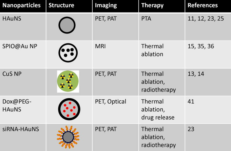
Nanomaterials that interact with light provide a unique opportunity for applications in biophotonic nanomedicine. Image-guided therapies could be designed based on multifunctional nanoparticles (NPs). Such NPs have a strong and tunable surface plasmon resonance absorption in the near-infrared region and can be detected using multiple imaging modalities (magnetic resonance imaging, nuclear imaging, and photoacoustic imaging). These novel nanostructures, once introduced, are expected to home in on solid tumors either via a passive targeting mechanism (i.e., the enhanced permeability and retention effect) or via an active targeting mechanism facilitated by ligands bound to their surfaces. Once the NPs reach their target tissue, their activity can then be turned on using an external stimulus. For example, photothermal conducting NPs primarily act by converting light energy into heat. As a result, the temperature in the treatment volume is elevated above the thermal damage threshold, which kills the cells. This process, termed photothermal ablation therapy (PTA), is effective, but it is also unlikely to kill all tumor cells when used alone. In addition to PTA, photothermal conducting NPs can also efficiently trigger the release of drugs and activate RNA interference. A multimodal approach, which permits simultaneous PTA therapy, chemotherapy, and therapeutic RNA interference, has the potential to completely eradicate residual diseased cells. In this Account, we provide an up-to-date review of the synthesis and characterization, functionalization, and in vitro and in vivo evaluation of NIR lightactivatable multifunctional nanostructures used for imaging and therapy. We emphasize research on hollow gold nanospheres, magnetic core-shell gold nanoshells, and semiconductor copper monosulfide NPs. We discuss three types of novel drug delivery systems in which hollow gold nanospheres are used to mediate controlled drug release.


Figures

FIGURE 1
FIGURE 1
Structures and functions of near-infrared light-activatable theranostic nanoparticles. HAuNSs, hollow gold nanospheres; SPIO, superparamagnetic iron oxide; DOX, doxorubicin; CuS, copper sulfide; NP, nanoparticle; PTA, photothermal ablation; RNAi, RNA interference; siRNA, small interfering RNA; PET, positron emission tomography; PAT, photoacoustic tomography; MRI, magnetic resonance imaging.
FIGURE 2
FIGURE 2
Selective PTA of A431 cells by C225-HAuNSs. (A) Cell viability after various treatments. Viable cells were stained green with calcein AM and dead cells were labeled red with EthD-1. Magnification, ×40. (B) Images of untreated viable cells and dead cells treated with C225-HAuNS and NIR laser at higher magnification (×400). The dead cells are shown by positive staining with EthD-1 (arrow, red). DIC, differential interference contrast. (Reproduced with permission from Ref. 12)
FIGURE 3
FIGURE 3
Biodistribution and intratumoral distribution of FITC-tagged HAuNSs. Tissue and tumors were removed 24 h after intravenous injection of HAuNSs. (A) Biodistribution data calculated as the number of particle aggregates per square millimeter of visual area at magnification ×200. Values are presented as mean ± SD (n = 5). *, P < 0.01. (B) Representative fluorescence micrographs of cryosectioned B16/F10 melanoma. Melanocortin type-1 receptor was stained with rabbit anti-mouse melanocortin type-1 receptor polyclonal antibody (pseudocolored red). Blood vessels were stained with rat anti-mouse CD31 monoclonal antibody (pseudocolored blue). NDP-MSH-PEG-HAuNSs are pseudocolored green. White arrowheads, melanocortin type-1 receptor. Asterisks, the lumens of tumor vasculature. (Reproduced with permission from Ref. 11)
FIGURE 4
FIGURE 4
(A) Representative T2*-weighted MRI scans of mice bearing A431 tumors before and after injection of SPIO@silica-Au NPs. (B) In vivo MRTI of a tumor injected intratumorally with SPIO@silica-Au NPs (top) and saline (bottom). At 24 h after injection of the agents, A431 tumors were irradiated with an 808-nm laser. Green arrows indicate the light path. Bar, 5 mm. (C) Temperature versus time for treatment volumes indicated by squares in (A), from 30 s before laser treatment (180 s, 4 W/cm2) until 90 s after laser treatment. Only in tumors injected with SPIO@silica-Au NPs was the temperature elevated above the 54°C threshold (dotted line) to ensure irreversible PTA of tumor cells. Arrows indicate when the laser was switched on and off. Values are presented as mean ± SD. (D) Representative plot of temperature versus depth below tumor surface. (Reproduced with permission from Ref. 36)
FIGURE 5
FIGURE 5
(A) MicroPET/CT image of nude mouse bearing subcutaneous U87 glioma xenograft, acquired at 24 h after intravenous injection of PEG-[64Cu]CuS NPs. Arrow: tumor. (B) CuS NPs induced PTA destruction of tumors in vivo at 24 h after NIR laser irradiation (12 W/cm2 at 808 nm for 5 min). i.v., intravenous; i.t., intratumoral. Values are presented as mean ± SD. Asterisks, p < 0.001 compared to saline only control. (Figure reproduced with permission from Ref. 14)
FIGURE 6
FIGURE 6
Schematic representation of the different NIR-activatable drug delivery systems.
FIGURE 7
FIGURE 7
(A) MicroPET/CT images of mice bearing subcutaneous HeLa cells after intravenous injection of 64Cu-FA-HAuNS-siRNANF-κB (left) and HAuNS-siRNANF-κB (control, right). Arrows: tumors. (B) NF-κB p65 expression in tumors exposed (top) and not exposed (bottom) to NIR laser. (Reproduced with permission from Ref. 23)

References

    1. Fiedler VU, Schwarzmaier HJ, Eickmeyer F, Muller FP, Schoepp C, Verreet PR. Laser-induced interstitial thermotherapy of liver metastases in an interventional 0. 5 Tesla MRI system: technique and first clinical experiences. J Magn Reson Imaging. 2001;13:729–737. - PubMed
    1. Overgaard J, Gonzalez Gonzalez D, Hulshof MC, Arcangeli G, Dahl O, Mella O, Bentzen SM. Randomised trial of hyperthermia as adjuvant to radiotherapy for recurrent or metastatic malignant melanoma. European Society for Hyperthermic Oncology. Lancet. 1995;345:540–543. - PubMed
    1. Sapareto SA, Dewey WC. Thermal dose determination in cancer therapy. Int J Radiat Oncol Biol Phys. 1984;10:787–800. - PubMed
    1. Rosenberg C, Puls R, Hegenscheid K, Kuehn J, Bollman T, Westerholt A, Weigel C, Hosten N. Laser ablation of metastatic lesions of the lung: long-term outcome. Am J Roentgenol. 2009;192:785–792. - PubMed
    1. Lindner U, Weersink RA, Haider MA, Gertner MR, Davidson SR, Atri M, Wilson BC, Fenster A, Trachtenberg J. Image guided photothermal focal therapy for localized prostate cancer: phase I trial. J Urol. 2009;182:1371–1377. - PubMed

Publication types