

Review
doi: 10.1089/AID.2011.0230.
Epub 2011 Sep 21.
HIV type 1 Gag as a target for antiviral therapy
Affiliations
- PMID: 21848364
- PMCID: PMC3251841
- DOI: 10.1089/AID.2011.0230
Item in Clipboard
Review
HIV type 1 Gag as a target for antiviral therapy
AIDS Res Hum Retroviruses.
2012 Jan.
Abstract
The Gag proteins of HIV-1 are central players in virus particle assembly, release, and maturation, and also function in the establishment of a productive infection. Despite their importance throughout the replication cycle, there are currently no approved antiretroviral therapies that target the Gag precursor protein or any of the mature Gag proteins. Recent progress in understanding the structural and cell biology of HIV-1 Gag function has revealed a number of potential Gag-related targets for possible therapeutic intervention. In this review, we summarize our current understanding of HIV-1 Gag and suggest some approaches for the development of novel antiretroviral agents that target Gag.
Figures
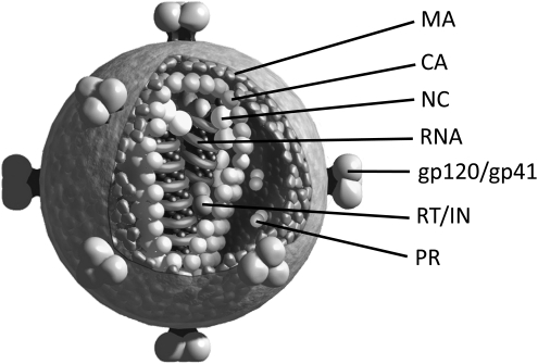
Representation of an HIV-1 virion. The locations of the Gag proteins matrix (MA), capsid (CA), and nucleocapsid (NC), the viral enzymes reverse transcriptase (RT), protease (PR), and integrase (IN), and the Env glycoproteins are indicated. The figure was generously provided by L. Henderson, National Cancer Institute, Frederick, MD.
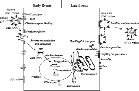
Schematic representation of the HIV-1 replication cycle. The details of the replication cycle are described in the text. Reprinted with permission from Freed (2004). Copyright 2004 Elsevier Inc.
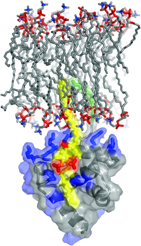
Model of HIV-1 MA binding to phosphatidylinositol-4,5-bisphosphate [PI(4,5)P2] in the membrane. The basic residues of MA (blue) interact electrostatically with phosphate groups (red) in PI(4,5)P2 (yellow and red). PI(4,5)P2 binding by MA induces exposure of the myristyl group (green), allowing it to insert into the membrane. The 2′-unsaturated acyl chain (yellow) of PI(4,5)P2 is predicted to bind to a hydrophobic cleft in MA. Unpublished image provided by M. Summers, based on the data of Saad et al. (2006). Color images available online at www.liebertonline.com/aid
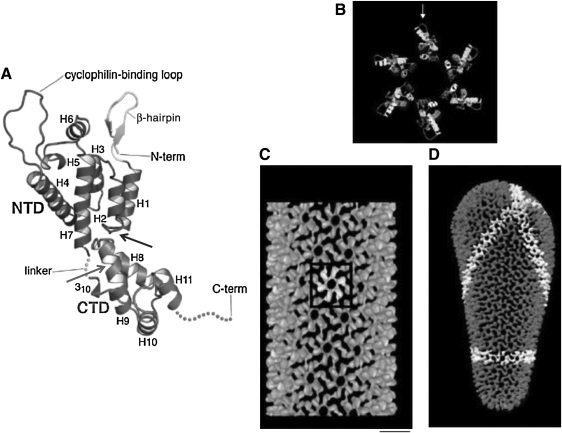
Structure of mature HIV-1 CA. (A) Tertiary structure of the mature monomeric CA with the NTD and CTD connected by the linker region. Helices 1–11 (H1–11), the N-terminal β-hairpin, and the cyclophilin A binding loop are shown. The C-terminal 11 residues of CA that are typically disordered in the crystal structure are indicated by a dashed line. The binding sites for CAI/NYAD-1/NYAD-13 and CAP1 are indicated by gray and black arrows, respectively. Reprinted with permission from Ganser-Pornillos et al. (2008). Copyright 2008 Elsevier Inc. (B) Molecular model of the hexameric ring formed by the NTD of CA, with the cyclophilin A-binding loop indicated with an arrow. (C) Outside view of the assembled tube structure, showing one hexamer (white) and the hexagonal CA lattice (gray). Scale bar=10 nm. (D) Model of an HIV-1 conical core. A continuous line of hexamers is highlighted in white. Reprinted with permission from Li et al. (2000). Copyright 2000 Macmillan Publishers Ltd: [Nature].
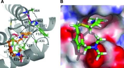
Structure of CAP-1 bound to the NTD of CA calculated by restrained molecular dynamics based on hybrid x-ray/NMR data. (A) View of CAP-1 bound to the NTD. The aromatic side-chain of Phe-32 displaced from the core upon CAP-1 binding is labeled and shown in green. (B) Representation of the electrostatic surface of CAP-1 bound to the NTD of CA. Upon CAP-1 binding, the aromatic ring of CAP-1 is inserted into the pocket formerly occupied by Phe-32. Adapted from Kelly et al. (2007). Copyright 2007 Elsevier, with permission. Color images available online at www.liebertonline.com/aid
Structure of the CAI peptide bound to a hydrophobic cavity of the CA CTD. The surface of the CTD is colored based on polarity: red, positively charged; blue, negatively charged; cyan, uncharged; white, nonpolar. The residues of CAI (yellow) in direct contact with the CTD, and N- and C-termini of CAI, are labeled. Reprinted from Ternois et al. (2005) with permission. Copyright 2005 Macmillan Publishers Ltd. [Nature Structural and Molecular Biology]. Color images available online at www.liebertonline.com/aid
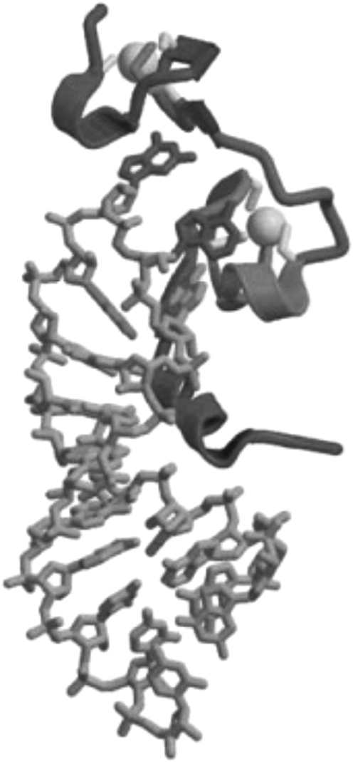
Structure of NC bound to the SL3 stem-loop of the HIV-1 RNA packaging signal. Gray balls represent the two zinc ions that bound to the zinc-finger motifs. Reprinted from Turner and Summers (1999) with permission. Copyright 1999 Elsevier Inc.
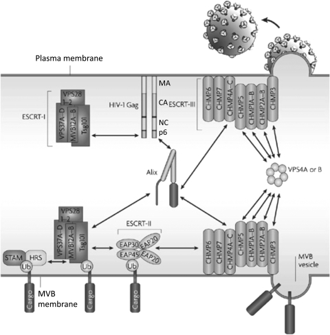
Schematic representation of the ESCRT machinery and its interaction with HIV-1 Gag at the plasma membrane and ubiquitinated cargo in the MVB. The MA, CA, NC, and p6 domains of Gag are shown. Double-headed arrows denote the interaction of p6 with Tsg101 and Alix, Alix with Tsg101, and Vps4 with ESCRT-III. A ubiquitinated (Ub) cargo protein is shown interacting with the STAM/Hrs complex (often referred to as ESCRT-0). Adapted with permission from Fujii et al. (2007). Copyright 2007 Macmillan Publishers Ltd. (Nature Reviews Microbiology).
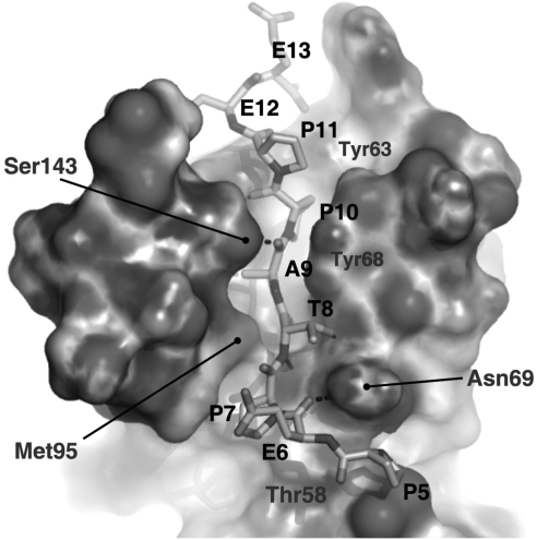
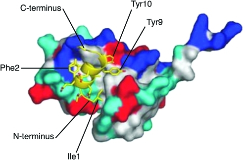
Structure of the UEV domain of Tsg101 bound to a PTAP-containing peptide derived from the sequence of HIV-1 p6. p6 residues Pro-5 (P5), Glu-6 (E6), Pro-7 (P7), Thr-8 (T8), Ala-9 (A9), Pro-10 (P10), Pro-11 (P11), Glu-12 (E12), and Glu-13 (E13) are shown, as are residues in Tsg101 that make contact with the PTAP-containing peptide. Reprinted with permission from Im et al. (2010). Copyright 2010 Cell Press.
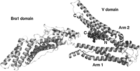
Structure of a YPXnL-containing peptide bound to the Alix V domain. Ribbon diagram of the Bro1 and V domain of ALIX in complex with the HIV-1 p6 YPXnL late-domain peptide. Adapted from Zhai et al. (2008). Copyright 2007 Macmillan Publishers Ltd. (Nature Structural and Molecular Biology).
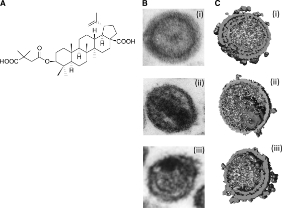
Bevirimat and its effect on HIV-1 maturation. (A) Structure of bevirimat. (B, C) Morphology of virions observed by thin-section EM (B) and cryoelectron tomography (C): (i) immature particles, (ii) mature particles, and (iii) particles produced from bevirimat-treated cells. Reprinted with permission from Adamson and Freed (2008) and Keller et al. (2011). Copyright 2008 Elsevier Inc. and 2011, American Society for Microbiology.
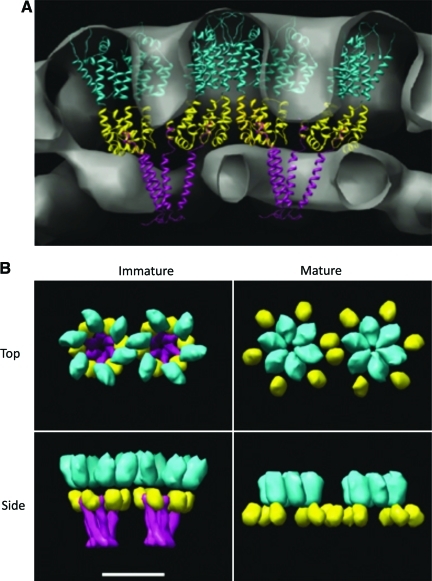
Model depicting the organization of CA in mature and immature Gag lattices. (A) Side view of the immature Gag lattice. The CA NTD (cyan), CA CTD (yellow), and SP1 (magenta) are shown. (B) Models representing the top and side views of Gag subunits in the immature (left) and mature (right) lattices. In the immature lattice, hexamers are held together by an SP1 bundle spaced by CTD interactions. SP1 is removed upon PR-mediated processing during maturation; in the mature lattice the hexamers are held together by the NTD spaced by CTD interactions. Scale bar=8 nm. Reprinted with permission from Wright et al. (2007). Copyright European Molecular Biology Organization. Color images available online at www.liebertonline.com/aid
References
-
- Berger EA. Murphy PM. Farber JM. Chemokine receptors as HIV‐1 coreceptors: Roles in viral entry, tropism, and disease. Annu Rev Immunol. 1999;17:657–700. - PubMed
-
- Doms RW. Beyond receptor expression: The influence of receptor conformation, density, and affinity in HIV-1 infection. Virology. 2000;276(2):229–237. - PubMed
-
- Freed EO. Martin MA. HIVs, their replication. In: Knipe DM, editor; Howley PM, editor. Fields Virology. Lippincott, Williams & Wilkins; Philadelphia: 2007. pp. 2107–2185.
Publication types
MeSH terms
Substances
Grants and funding
LinkOut - more resources
Full Text Sources
Other Literature Sources
Medical
