. 2011 Sep;81(6):1511-25.
doi: 10.1111/j.1365-2958.2011.07787.x. Epub 2011 Aug 17.

Inactivation of a Plasmodium apicoplast protein attenuates formation of liver merozoites

Affiliations
Free PMC article

Inactivation of a Plasmodium apicoplast protein attenuates formation of liver merozoites

Joana M Haussig et al. Mol Microbiol. 2011 Sep.
Free PMC article

Abstract

Malaria parasites undergo a population expansion inside the host liver before disease onset. Developmental arrest inside host hepatocytes elicits protective immune responses. Therefore, elucidation of the molecular mechanisms leading to mature hepatic merozoites, which initiate the pathogenic blood phase, also informs anti-malaria vaccine strategies. Using targeted gene deletion in the rodent model malaria parasite Plasmodium berghei, we show that a Plasmodium-specific Apicoplast protein plays an important role for Liver Merozoite formation (PALM). While the resulting knockout mutants develop normally for most of the life cycle, merozoite release into the blood stream and the ability to establish an infection are severely impaired. Presence of a signature blood-stage antigen, merozoite surface protein 1 and normal apicoplast morphology indicate that the inability to finalize merozoite segregation is a direct consequence of loss of PALM function. Experimental immunization of mice with as few as two doses of palm(-) sporozoites can elicit sterile protection up to 110 days after final immunization. Our data establish that a tailor-made arrest in the final steps of hepatic merozoite formation can induce strong protective immune responses and that malaria parasites employ a distinct apicoplast protein for efficient formation of pre-erythrocytic merozoites.


Figures

Fig. 1
Fig. 1
The Plasmodium-specific apicoplast protein important for liver merozoite formation (PALM). A. Primary structure of Plasmodium PALM proteins. Shown are the overall sequence structures and amino acid sequence identities of PALM orthologues in P. yoelii (PY01863), P. falciparum (PFF0110w) and P. vivax (PVX_113280), compared with P. berghei PALM (PBANKA_010110). Signal peptide (red), apicoplast-targeting sequence (green) as predicted with PlasmoAP (Foth et al., 2003), and the conserved domain (grey) with two signature cysteine residues (yellow) are shown. B. Sequence alignment of Plasmodium PALM proteins. Shown is the conserved domain (A) with the two signature cysteine residues (highlighted yellow). P. berghei PALM, PBANKA_010110; P. yoelii PALM, PY01863; P. chabaudi PALM, PCHAS_010180; P. falciparum PALM, PFF0110w; P. vivax PALM, PVX_113280; P. knowlesi PALM, PKH_114760.
Fig. 2
Fig. 2
Live cell imaging of PALM in infected hepatoma cells. A. Generation of PALM-mCherry-myc parasites. The PbPALM genomic locus was targeted with a replacement plasmid containing the C-terminal PALM fragment (grey) fused in-frame to the mCherry coding sequence (red), and a quadruple c-myc tag (blue) and followed by the 3′UTR of PbDHFS/FPGS. In addition, the targeting plasmid contains the PbDHFR/TS positive selectable marker (black box), and a fragment of the PALM 3′UTR. Upon a double cross-over event, the targeting plasmid is expected to replace the endogenous PALM ORF with a C-terminally tagged PALM fusion protein. Arrows and bars indicate specific primers and PCR fragments respectively. B. Genotyping of the PALM-mCherry-myc parasite line. Using integration-specific primer combinations (A), the successful replacement event was verified. Absence of the WT signal from PALM-mCherry-myc parasites confirmed the purity of the clonal population. C. Hepatoma cells were infected with PALM-mCherry-myc sporozoites. PALM expression was visualized in late liver stages 2 days after infection. Note the branched structure of the mCherry signal. Bars, 10 µm.
Fig. 3
Fig. 3
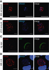
Expression of PALM during the Plasmodium berghei life cycle. PALM-mCherry-myc parasites were used to infect mice and Anopheles stephensi mosquitoes. Intra- and extracellular parasite stages were fixed, permeabilized and stained with mouse anti-myc antibody (red). In replicating stages, nuclei were stained with the DNA-dye DRAQ5 (blue). The GFP signal in fixed sporozoites was used to display the extracellular parasite. A. Blood-stage schizont (BS schizont) from an infected mouse. Note that the anti-myc signal is very weak and was only detectable after background subtraction in comparison with WT parasite-infected erythrocytes. Bars, 5 µm. B. Oocysts fixed at day 10 after infection. Bars, 10 µm. C. Salivary gland sporozoites fixed at day 17 after infection. Bars, 10 µm. D. Liver-stage parasites fixed at 48 h after infection of hepatoma cells. Bars, 10 µm. Note the branched structure indicative of an apicoplast localization of PALM.
Fig. 4
Fig. 4
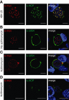
Apicoplast localization of PALM. A. Co-staining of fixed, PALM-mCherry-myc parasite-infected hepatoma cells 48 h after infection using anti-myc and anti-ACP antibodies. Note the substantial overlap between PALM and the signature apicoplast protein. B. PALM-mCherry-myc parasite-infected hepatoma cells left untreated, fixed at 48 h after infection, and stained with antibodies specific for the myc epitope and upregulated in infectious sporozoites protein 4 (UIS4), a signature protein of the parasitophorous vacuole. Note the branched PALM-positive structures. C. Antibiotics treatment with 1 µM azithromycin abolishes the branched PALM-positive structures while parasites appear to remain healthy otherwise. D. Antibiotics treatment with 1 µM azithromycin abolishes the branched PALM-positive structures as well as ACP-positive structures. All bars, 10 µm.
Fig. 5
Fig. 5
Generation of palm- parasites. A. The PbPALM genomic locus was targeted with a replacement plasmid containing PALM 5′ and 3′ fragments (thick black lines) and the DHFR/TS positive selectable marker (black box). Upon a double cross-over event, the targeting plasmid is expected to replace the endogenous PALM ORF. Arrows and light grey solid lines indicate specific primers and PCR fragments respectively. Black bars indicate specific 5′ and 3′ probes; dark grey dashed lines represent EcoRI or KpnI restriction-digested fragments of WT or knockout parasite gDNA. B. Mouse-mosquito-mouse transmission experiment starting from a mixed WT/palm- infection (input) and resulting in a pure WT population after completion of one transmission cycle (output). Parasites were genotyped by PCR using wild-type (WT)- and replacement (INT)-specific primer pairs, as indicated in (A). C. Alternative gene replacement vector to ablate the PALM locus. The PbPALM genomic locus was targeted with a replacement plasmid containing a PALM 5′ fragment, containing a portion of the PALM open reading frame (grey box), fused to the mCherry protein (red box) and quadruple myc tag (blue box), followed by the DHFR/TS positive selectable marker (black box) and the 3′ flanking region (thick bar). Upon a double cross-over event, the targeting plasmid is expected to replace the endogenous PALM ORF. Arrows and bars indicate specific primers and PCR fragments respectively. D. Genotyping of the palm- ANKA-GFP parasite line used for the majority of experiments through Southern blot analysis. Probes at the 5′ and 3′ UTR of PALM were used to demonstrate the expected size shift of restriction-digested gDNA of WT and knockout parasites. E. Genotyping of the three clonal palm- parasite lines obtained from independent transfection experiments. Using integration-specific primer combinations (A, C) the successful replacement event (INT) was verified. Absence of the WT signal from palm- parasites confirmed the purity of the clonal populations. The clonal populations palm- ANKA (blue) and palm- ANKA-GFP (green) were obtained according to design (A), using WT-ANKA and ANKA-GFP parasites as recipient lines (left); the clonal population palm--NT-tag (red) was obtained according to design (C), using ANKA-GFP parasites as recipient lines (right).
Fig. 6
Fig. 6
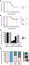
Infection with palm- sporozoites leads to attenuated liver-stage development in vivo. A. Kaplan–Meier analysis of time to malaria blood-stage infection. C57BL/6 mice were infected by natural bite with five mosquitoes infected with ANKA-GFP (grey, n = 11) or palm- ANKA-GFP (black, n = 13). Animals were monitored daily for presence of parasites in Giemsa-stained blood smears. B. Kaplan–Meier analysis of time to malaria blood-stage infection. C57BL/6 mice were infected with 1000, 10 000, or 100 000 ANKA-GFP (blue, n = 3, 9, 3) or palm- ANKA-GFP (red, n = 20, 82, 45) sporozoites by intravenous injection. Animals were monitored daily for presence of parasites in Giemsa-stained blood smears. C. Development of symptoms of ECM is only reduced in palm- sporozoite-infected animals. Shown is the percentage of parasite-positive C57BL/6 mice that develop signature symptoms of ECM (i.e. mice showing sudden onset of ataxia, paralysis, convulsion or coma, or a minimum of three behavioural and functional abnormalities) after injection of 1000 blood-stage parasites (ANKA-GFP, grey, n = 15; palm- ANKA-GFP, black, n = 15), injection of sporozoites [10 000 ANKA-GFP, grey, n = 9; palm- ANKA-GFP, black, 1000 (n = 6), 10 000 (n = 20), or 100 000 (n = 13)], or infection by natural bite with five mosquitoes infected with ANKA-GFP (grey, n = 11) or palm- ANKA-GFP (black, n = 13). D. Integrity of the blood–brain barrier is preserved in most palm- sporozoite-infected mice. Mice were infected with 10 000 ANKA-GFP sporozoites (n = 4), or 10 000 (n = 5) or 100 000 (n = 7) palm- ANKA-GFP sporozoites. Evan's blue was injected intravenously at day 4 after appearance of blood-stage parasites and brains removed for documentation. One mouse infected with 100 000 palm- ANKA-GFP sporozoites showed behavioural abnormalities and breakdown of the blood–brain barrier.
Fig. 7
Fig. 7
palm- parasites display a defect in liver-stage maturation. A. Quantification of liver stages in cultured hepatoma cells at 24, 48 and 72 h after infection with 10 000 P. berghei ANKA-GFP and palm- ANKA-GFP sporozoites. Infected cells were fixed and stained with mouse anti-PbHSP70 antibodies. Note that in contrast to ANKA-GFP parasites, palm- ANKA-GFP parasites remain inside the host cells even at 72 h after infection. Data are from two independent experiments done in triplicate. Shown are mean numbers (± SD). B. Parasite transcript detection in WT and palm- parasite-infected livers confirms the successful ablation of PALM in the knockout line. cDNA was prepared from liver homogenates extracted from mice at 44 h after sporozoite infection. Quality of cDNA preparations and presence of parasites in vivo was controlled using HSP70-specific primers. C. Defect of merosome formation in palm- parasites. Quantification of merosomes obtained from cultured hepatoma cells infected with ANKA-GFP and palm- ANKA-GFP parasites at 72 h after inoculation with 100 000 sporozoites. D. palm- ANKA-GFP merosomes are infectious to mice. Complete merosome containing supernatants harvested under (C) were injected into recipient C57BL/6 mice (ANKA-GFP, n = 5; palm-, n = 4) and monitored for malaria infections by examination of Giemsa-stained blood-smears. E. Apicoplast morphology appears normal in palm- liver stages. Hepatoma cells infected with WT and palm- parasites were fixed at 48 or 60 h after infection and stained for parasite cytoplasm and the apicoplast using anti-HSP70 and anti-ACP antibodies respectively. The merge includes an additional DRAQ5 nuclear stain. Bars, 10 µm.
Fig. 8
Fig. 8
palm- parasites cannot finalize liver merozoite formation efficiently. A. Defect in liver-stage merozoite segregation in palm- parasites. In vitro cultured P. berghei ANKA-GFP and palm- ANKA-GFP liver stages, were fixed at various time points after sporozoite infection and visualized by immunofluorescence using monoclonal mouse anti-P. yoelii MSP1 and rabbit anti-P. berghei UIS4 antibodies. Nuclei were stained with the DNA-dye DRAQ5 (blue). palm- ANKA-GFP parasites develop normally until late liver stages but rarely form merozoites. Shown are representative images of ANKA-GFP and palm- ANKA-GFP at various time points. Bars, 10 µm. B. Quantification of productive liver merozoite formation. Liver stages from ANKA-GFP and palm- ANKA-GFP parasites were scored at 48, 60 and 72 h after infection (ANKA-GFP n = 105, 94, 82; palm-n = 99, 124, 107) according to the five categories indicated by the representative images in the inset. The categories are: no MSP1 (white), rim staining of MSP1 (light green), intracellular schizont staining of MSP1 (dark green), formation of MSP1-positive merozoites (yellow), and aberrant, external MSP1 staining (black). Note the high proportion of MSP1-positive schizonts in palm- parasites, which correlates with a low production of MSP1-positive pathogenic merozoites.

Similar articles

Cited by

References

    1. Amino R, Thiberge S, Martin B, Celli S, Shorte S, Frischknecht F, Ménard R. Quantitative imaging of Plasmodium transmission from mosquito to mammal. Nat Med. 2006;12:220–224. - PubMed
    1. Baer K, Klotz C, Kappe SHI, Schnieder T, Frevert U. Release of hepatic Plasmodium yoelii merozoites into the pulmonary microvasculature. PLoS Pathog. 2007;3:e171. - PMC - PubMed
    1. Chitale M, Hawkins T, Park C, Kihara D. ESG: extended similarity group method for automated protein function prediction. Bioinformatics. 2009;25:1739–1745. - PMC - PubMed
    1. Cohen J, Nussenzweig V, Nussenzweig R, Vekemans J, Leach A. From the circumsporozoite protein to the RTS, S/AS candidate vaccine. Hum Vaccin. 2010;6:90–96. - PubMed
    1. Dahl EL, Rosenthal PJ. Multiple antibiotics exert delayed effects against the Plasmodium falciparum apicoplast. Antimicrob Agents Chemother. 2007;51:3485–3490. - PMC - PubMed

Publication types

MeSH terms