

Reduced functional loads alter the physical characteristics of the bone-periodontal ligament-cementum complex
- PMID: 21848615
- PMCID: PMC3200425
- DOI: 10.1111/j.1600-0765.2011.01396.x
Reduced functional loads alter the physical characteristics of the bone-periodontal ligament-cementum complex
Abstract
Background and objective: Adaptive properties of the bone-periodontal ligament-tooth complex have been identified by changing the magnitude of functional loads using small-scale animal models, such as rodents. Reported adaptive responses as a result of lower loads due to softer diet include decreased muscle development, change in structure-function relationship of the cranium, narrowed periodontal ligament space, and changes in the mineral level of the cortical bone and alveolar jaw bone and in the glycosaminoglycans of the alveolar bone. However, the adaptive role of the dynamic bone-periodontal ligament-cementum complex to prolonged reduced loads has not been fully explained to date, especially with regard to concurrent adaptations of bone, periodontal ligament and cementum. Therefore, in the present study, using a rat model, the temporal effect of reduced functional loads on physical characteristics, such as morphology and mechanical properties and the mineral profiles of the bone-periodontal ligament-cementum complex was investigated.
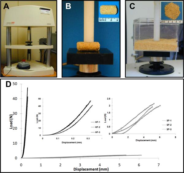
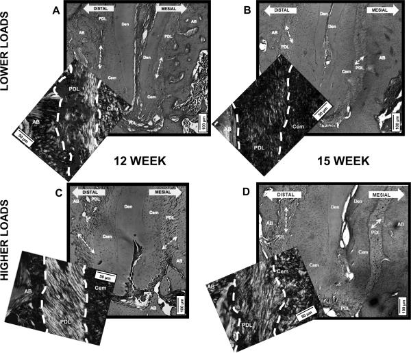
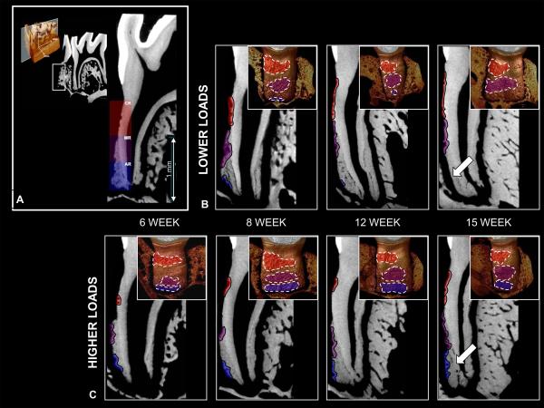
Material and methods: Two groups of 6-wk-old male Sprague-Dawley rats were fed nutritionally identical food with a stiffness range of 127-158 N/mm for hard pellet or 0.3-0.5 N/mm for soft powder forms. Spatio-temporal adaptation of the bone-periodontal ligament-cementum complex was identified by mapping changes in the following: (i) periodontal ligament collagen orientation and birefringence using polarized light microscopy, bone and cementum adaptation using histochemistry, and bone and cementum morphology using micro-X-ray computed tomography; (ii) mineral profiles of the periodontal ligament-cementum and periodontal ligament-bone interfaces by X-ray attenuation; and (iii) microhardness of bone and cementum by microindentation of specimens at ages 6, 8, 12 and 15 wk.
Results: Reduced functional loads over prolonged time resulted in the following adaptations: (i) altered periodontal ligament orientation and decreased periodontal ligament collagen birefringence, indicating decreased periodontal ligament turnover rate and decreased apical cementum resorption; (ii) a gradual increase in X-ray attenuation, owing to mineral differences, at the periodontal ligament-bone and periodontal ligament-cementum interfaces, without significant differences in the gradients for either group; (iii) significantly (p < 0.05) lower microhardness of alveolar bone (0.93 ± 0.16 GPa) and secondary cementum (0.803 ± 0.13 GPa) compared with the higher load group insert bone = (1.10 ± 0.17 and cementum = 0.940 ± 0.15 GPa, respectively) at 15 wk, indicating a temporal effect of loads on the local mineralization of bone and cementum.
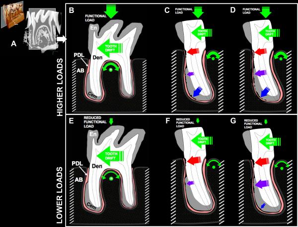
Conclusion: Based on the results from this study, the effect of reduced functional loads for a prolonged time could differentially affect morphology, mechanical properties and mineral variations of the local load-bearing sites in the bone-periodontal ligament-cementum complex. These observed local changes in turn could help to explain the overall biomechanical function and adaptations of the tooth-bone joint. From a clinical translation perspective, our study provides an insight into modulation of load on the complex for improved tooth function during periodontal disease and/or orthodontic and prosthodontic treatments.
© 2011 John Wiley & Sons A/S.
Figures


Similar articles
-
Multiscale biomechanical responses of adapted bone-periodontal ligament-tooth fibrous joints.Bone. 2015 Dec;81:196-207. doi: 10.1016/j.bone.2015.07.004. Epub 2015 Jul 4. Bone. 2015. PMID: 26151121 Free PMC article.
-
Biomechanical pathways of dentoalveolar fibrous joints in health and disease.Periodontol 2000. 2020 Feb;82(1):238-256. doi: 10.1111/prd.12306. Periodontol 2000. 2020. PMID: 31850635 Review.
-
The biomechanical characteristics of the bone-periodontal ligament-cementum complex.Biomaterials. 2010 Sep;31(25):6635-46. doi: 10.1016/j.biomaterials.2010.05.024. Epub 2010 Jun 11. Biomaterials. 2010. PMID: 20541802 Free PMC article.
-
Differentiating zones at periodontal ligament-bone and periodontal ligament-cementum entheses.J Periodontal Res. 2015 Dec;50(6):870-80. doi: 10.1111/jre.12281. Epub 2015 Jun 1. J Periodontal Res. 2015. PMID: 26031604 Free PMC article.
-
A Force on the Crown and Tug of War in the Periodontal Complex.J Dent Res. 2018 Mar;97(3):241-250. doi: 10.1177/0022034517744556. Epub 2018 Jan 24. J Dent Res. 2018. PMID: 29364757 Free PMC article. Review.
Cited by
-
High Frequency Acceleration: A New Tool for Alveolar Bone Regeneration.JSM Dent Surg. 2017;2(4):1026. Epub 2017 Aug 25. JSM Dent Surg. 2017. PMID: 30215055 Free PMC article.
-
[Research progress on labial protuberances of anterior teeth in orthodontic treatment].Zhejiang Da Xue Xue Bao Yi Xue Ban. 2024 Oct 25;53(5):586-592. doi: 10.3724/zdxbyxb-2024-0019. Zhejiang Da Xue Xue Bao Yi Xue Ban. 2024. PMID: 39183065 Free PMC article. Review. Chinese.
-
Mechanoadaptive strain and functional osseointegration of dental implants in rats.Bone. 2020 Aug;137:115375. doi: 10.1016/j.bone.2020.115375. Epub 2020 Apr 23. Bone. 2020. PMID: 32335376 Free PMC article.
-
Multifactorial analysis of factors influencing premolar mobility in stage III/IV grade C periodontitis patients ≤ 35 years of age: a cross-sectional study.BMC Oral Health. 2024 Oct 16;24(1):1232. doi: 10.1186/s12903-024-05039-2. BMC Oral Health. 2024. PMID: 39415252 Free PMC article.
-
Chewing-Activated TRPV4/PIEZO1-HIF-1α-Zn Axes in a Rat Periodontal Complex.J Dent Res. 2025 Apr;104(4):398-407. doi: 10.1177/00220345241294001. Epub 2025 Jan 28. J Dent Res. 2025. PMID: 39876056 Free PMC article.
References
-
- Carter DR, Beaupre GS. Skeletal function and form. Cambridge University Press; 2001.
-
- Nanci A, Bosshardt DD. Structure of periodontal tissues in health and disease. Periodontol 2000. 2006;40:11–28. - PubMed
-
- Ten Cate AR. Oral Histology Development, Structure, and Function. 5th edn. Mosby Year Book Inc.; 1998.
-
- Woda A, Foster K, Mishellany A, Peyron MA. Adaptation of healthy mastication to factors pertaining to the individual or to the food. Physiol Behav. 2006;89:28–35. - PubMed
-
- Graf H. In: Occlusal forces and mandibular movements. Elmsford NY, editor. Pergamon Press, Inc.; 1978.
Publication types
MeSH terms
Substances
Grants and funding
LinkOut - more resources
Full Text Sources
