

The muscle satellite cell at 50: the formative years
- PMID: 21849021
- PMCID: PMC3177780
- DOI: 10.1186/2044-5040-1-28
The muscle satellite cell at 50: the formative years
Abstract
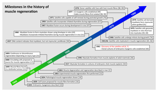
In February 1961, Alexander Mauro described a cell 'wedged' between the plasma membrane of the muscle fibre and the surrounding basement membrane. He postulated that it could be a dormant myoblast, poised to repair muscle when needed. In the same month, Bernard Katz also reported a cell in a similar location on muscle spindles, suggesting that it was associated with development and growth of intrafusal muscle fibres. Both Mauro and Katz used the term 'satellite cell' in relation to their discoveries. Today, the muscle satellite cell is widely accepted as the resident stem cell of skeletal muscle, supplying myoblasts for growth, homeostasis and repair.Since 2011 marks both the 50th anniversary of the discovery of the satellite cell, and the launch of Skeletal Muscle, it seems an opportune moment to summarise the seminal events in the history of research into muscle regeneration. We start with the 19th-century pioneers who showed that muscle had a regenerative capacity, through to the descriptions from the mid-20th century of the underlying cellular mechanisms. The journey of the satellite cell from electron microscope curio, to its gradual acceptance as a bona fide myoblast precursor, is then charted: work that provided the foundations for our understanding of the role of the satellite cell. Finally, the rapid progress in the age of molecular biology is briefly discussed, and some ongoing debates on satellite cell function highlighted.
Figures


References
-
- Janssen I, Heymsfield SB, Wang ZM, Ross R. Skeletal muscle mass and distribution in 468 men and women aged 18-88 yr. J Appl Physiol. 2000;89:81–88. - PubMed
-
- Zenker FA. Über die Veraenderungen der willkührlichen Muskeln im Typhus abdominalis. Leipzig, Germany: Vogel; 1864.
-
- Waldeyer W. Ueber die Veränderungen der quergestreiften Muskeln bei der Entzündung und dem Typhusprozess, sowie über die Regeneration derselben nach Substanzdefecten. Virchows Archiv. 1865;34:472–514. doi: 10.1007/BF02323030. - DOI
-
- Weber CO. Ueber die Neubildung quergestreifter Muskelfasern, insbesondere die regenerative Neubildung derselben nach Verletzungen. Virchows Archiv. 1867;39:216–253. doi: 10.1007/BF01879135. - DOI
-
- Weber CO. Ueber die Betheiligung der Muskelkörperchen und der quergestreiften Muskeln an den Neubildungen nebst Bemerkungen über die Lehre von der Specificität der Gewebselemente. Virchows Archiv. 1867;39:254–269. doi: 10.1007/BF01879136. - DOI
LinkOut - more resources
Full Text Sources
