. 2011 Aug 17:6:96.
doi: 10.1186/1748-717X-6-96.

Mechanisms of increased risk of tumorigenesis in Atm and Brca1 double heterozygosity

Affiliations

Mechanisms of increased risk of tumorigenesis in Atm and Brca1 double heterozygosity

Jufang Wang et al. Radiat Oncol. .

Abstract

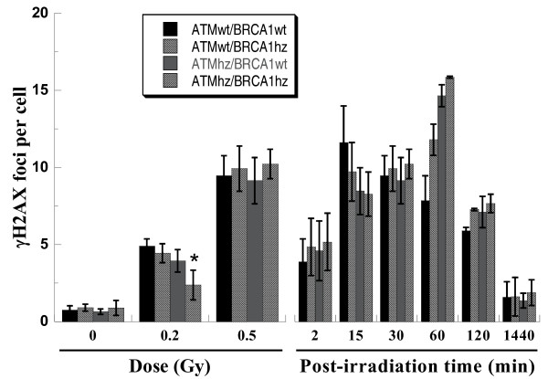
Background: Both epidemiological and experimental studies suggest that heterozygosity for a single gene is linked with tumorigenesis and heterozygosity for two genes increases the risk of tumor incidence. Our previous work has demonstrated that Atm/Brca1 double heterozygosity leads to higher cell transformation rate than single heterozygosity. However, the underlying mechanisms have not been fully understood yet. In the present study, a series of pathways were investigated to clarify the possible mechanisms of increased risk of tumorigenesis in Atm and Brca1 heterozygosity.

Methods: Wild type cells, Atm or Brca1 single heterozygous cells, and Atm/Brca1 double heterozygous cells were used to investigate DNA damage and repair, cell cycle, micronuclei, and cell transformation after photon irradiation.

Results: Remarkable high transformation frequency was confirmed in Atm/Brca1 double heterozygous cells compared to wild type cells. It was observed that delayed DNA damage recognition, disturbed cell cycle checkpoint, incomplete DNA repair, and increased genomic instability were involved in the biological networks. Haploinsufficiency of either ATM or BRCA1 negatively impacts these pathways.

Conclusions: The quantity of critical proteins such as ATM and BRCA1 plays an important role in determination of the fate of cells exposed to ionizing radiation and double heterozygosity increases the risk of tumorigenesis. These findings also benefit understanding of the individual susceptibility to tumor initiation.


Figures

Figure 1
Figure 1
Anchorage-independent growth levels of four kinds of MEF cells. After cells were exposed to 2 Gy of photons, permissive soft-agar assay was performed immediately to assess cell survival (Panel A) and 1 week later, non-permissive soft-agar assay were performed to assess neoplastic transformation (Panel B). Transformants were visually scored under a dissecting microscope. Data are from four independent experiments. * indicates the significant difference (p < 0.05) compared with ATMwt/BRCA1wt cells and ^ indicates the significant difference (p < 0.05) compared with ATMwt/BRCA1hz cells experienced the same treatment.
Figure 2
Figure 2
Representative pictures of γH2AX foci. Cells were exposed to 0.5 Gy of photons and stained with mouse anti-γH2AX antibody visualized by Alexa Fluor® 488 anti-mouse antibody and propidium iodide (PI) at 15 and 1440 min post-irradiation.
Figure 3
Figure 3
Quantitative detection of DSBs by counting γH2AX foci. Dose response (left) and kinetics (right) of γH2AX focus formation. Samples for dose response were fixed at 30 min after irradiation and kinetics was for 0.5Gy of photons. * indicates the significant difference (p < 0.05) compared with ATMwt/BRCA1wt cells exposed to 0.2 Gy of photons.
Figure 4
Figure 4
Total DNA damage measured with alkaline comet assay in four kinds of MEF cells at different time course. Data were a pool of three independent experiments and totally 200 cells were scored. * and ** indicate the significant difference compared with ATMwt/BRCA1wt cells experienced the same treatment and same post-incubation (* for p < 0.05 and ** p < 0.01).
Figure 5
Figure 5
Cell cycle distribution in MEF cells exposed to 5 Gy of photons. Kinetics of G2/M block based on original data (Panel A) and optimized dada in which the control level of G2/M-phase cells was correspondingly subtracted from the proportion of G2/M-phase cells of each sample (Panel B). Data are pooled from three independent experiments.
Figure 6
Figure 6
Induction of micronuclei in MEF cells exposed to 2 Gy of photons. Individual cells were visualized with Acridine Orange staining and micronuclei (MN) per binucleated cell (BN) were score under fluorescence microscope. More than 500 BN cells were counted for each sample. Data were pooled from three independent experiments. * indicates the significant difference (p < 0.05) compared with ATMwt/BRCA1wt cells exposed to the same dose of photons.

References

    1. Kwon CH, Zhao D, Chen J, Alcantara S, Li Y, Burns DK, Mason RP, Lee EY, Wu H, Parada LF. Pten haploinsufficiency accelerates formation of high-grade astrocytomas. Cancer Res. 2008;68:3286–3294. doi: 10.1158/0008-5472.CAN-07-6867. - DOI - PMC - PubMed
    1. Le Toriellec E, Despouy G, Pierron G, Gaye N, Joiner M, Bellanger D, Vincent-Salomon A, Stern MH. Haploinsufficiency of CDKN1B contributes to leukemogenesis in T-cell prolymphocytic leukemia. Blood. 2008;111:2321–2328. doi: 10.1182/blood-2007-06-095570. - DOI - PubMed
    1. Kucherlapati M, Yang K, Kuraguchi M, Zhao J, Lia M, Heyer J, Kane MF, Fan K, Russell R, Brown AM, Kneitz B, Edelmann W, Kolodner RD, Lipkin M, Kucherlapati R. Haploinsufficiency of Flap endonuclease (Fen1) leads to rapid tumor progression. Proc Natl Acad Sci USA. 2002;99:9924–9929. doi: 10.1073/pnas.152321699. - DOI - PMC - PubMed
    1. Kleiman NJ, David J, Elliston CD, Hopkins KM, Smilenov LB, Brenner DJ, Worgul BV, Hall EJ, Lieberman HB. Mrad9 and atm haploinsufficiency enhance spontaneous and X-ray-induced cataractogenesis in mice. Radiat Res. 2007;168:567–573. doi: 10.1667/rr1122.1. - DOI - PubMed
    1. Izeradjene K, Combs C, Best M, Gopinathan A, Wagner A, Grady WM, Deng CX, Hruban RH, Adsay NV, Tuveson DA, Hingorani SR. Kras(G12D) and Smad4/Dpc4 haploinsufficiency cooperate to induce mucinous cystic neoplasms and invasive adenocarcinoma of the pancreas. Cancer Cell. 2007;11:229–243. doi: 10.1016/j.ccr.2007.01.017. - DOI - PubMed

Publication types

MeSH terms