

Case Reports
doi: 10.1259/bjr/95083086.
Not the typical Tornwaldt's cyst this time? A nasopharyngeal cyst associated with canalis basilaris medianus
Affiliations
- PMID: 21849356
- PMCID: PMC3473778
- DOI: 10.1259/bjr/95083086
Item in Clipboard
Case Reports
Not the typical Tornwaldt's cyst this time? A nasopharyngeal cyst associated with canalis basilaris medianus
Br J Radiol.
2011 Sep.
Abstract
We report a patient with a cystic structure in the nasopharynx mimicking a Tornwaldt's cyst, which was felt to represent a different entity owing to the lack of the distinct features of a typical Tornwaldt's cyst. It was associated with a bony cleft in the basiocciput that was considered to be a canalis basilaris medianus (CBM), thought to represent an embryological vestige of the cephalic end of the notochord along its course within the basiocciput.
Figures

(a–d) Axial fat saturated T2 weighted images. (a) Image through the cystic lesion (arrows) in the posterior midline nasopharnyx. (b) Slice more superiorly demonstrating the defect in the clivus. Arrows denote the partially hyperintense defect in the midline. (c) Inverted T2 weighted image through the same level as (b) with better depiction of the canal within the bone. (d) More superior slice demonstrating the junction of the canal with the dural surface. (e and f) Thin slice CT images. (e) Axial and (f) sagittal images thin images from a dedicated skull base CT depicting the bony canal within the clivus. Arrow in (e) depict the broader anterior part of the canal which could be a so-called “foveola pharyngica”. Arrowheads in (e) point to the sclerotic posterior aspect of the defect. Arrows in (f) denote the broader anterior aspect; cranial communication could not be definitively demonstrated.
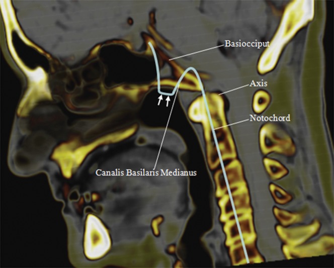
A different, normal patient with a sagittal view of the skull base showing a superimposed diagram of the craniocervical component of the expected embryonic notochordal trajectory in the adult (coloured line). The cephalic end of the notochord may be embedded in the bony sphenoid, while the segment in the basocciput level (arrows) may lie free in the nasopharyngeal wall.
Similar articles
-
Tornwaldt's cyst presenting only as occipital headache: a case report.Headache. 2009 Feb;49(2):307-10. doi: 10.1111/j.1526-4610.2008.01186.x. Epub 2008 Jul 3. Headache. 2009. PMID: 18631187
-
[MR imaging of the Tornwaldt's cyst: retrospective study of 1300 patients].Recenti Prog Med. 2013 Jul-Aug;104(7-8):398-402. doi: 10.1701/1315.14583. Recenti Prog Med. 2013. PMID: 24042415 Italian.
-
Tornwaldt's bursa.Clin Otolaryngol Allied Sci. 1985 Feb;10(1):21-5. doi: 10.1111/j.1365-2273.1985.tb01160.x. Clin Otolaryngol Allied Sci. 1985. PMID: 4006264
-
Tornwaldt's disease.Acta Otolaryngol Suppl. 1994;517:36-9. doi: 10.3109/00016489409124336. Acta Otolaryngol Suppl. 1994. PMID: 7856446 Review.
-
[Nasopharyngeal cysts. Report of four cases and literature review].An Otorrinolaringol Ibero Am. 1999;26(6):607-19. An Otorrinolaringol Ibero Am. 1999. PMID: 10645019 Review. Spanish.
Cited by
-
Pharyngeal enterogenous cyst associated with canalis basilaris medianus in a newborn.Pediatr Radiol. 2013 Apr;43(4):512-5. doi: 10.1007/s00247-012-2513-0. Epub 2012 Nov 9. Pediatr Radiol. 2013. PMID: 23138685
-
Nasopharyngeal cyst in an adolescent boy: differential diagnosis - embryological and anatomical considerations.BMJ Case Rep. 2023 Nov 22;16(11):e256945. doi: 10.1136/bcr-2023-256945. BMJ Case Rep. 2023. PMID: 37993143
-
Neuroradiological findings in Alagille syndrome.Br J Radiol. 2022 Jan 1;95(1129):20201241. doi: 10.1259/bjr.20201241. Epub 2021 Oct 5. Br J Radiol. 2022. PMID: 34609904 Free PMC article. Review.
-
Radiologic evaluation of the fossa navicularis: incidence, morphometric features, and clinical implications.Surg Radiol Anat. 2021 Nov;43(11):1887-1893. doi: 10.1007/s00276-021-02742-5. Epub 2021 Apr 16. Surg Radiol Anat. 2021. PMID: 33860857
-
Evaluation of canalis basilaris medianus using cone-beam computed tomography.Imaging Sci Dent. 2016 Jun;46(2):141-4. doi: 10.5624/isd.2016.46.2.141. Epub 2016 Jun 23. Imaging Sci Dent. 2016. PMID: 27358822 Free PMC article.
References
-
- Gruber W. Over the canalis basilaris medianus anomalies of the occipital bone in humans. Mem Acad Imp Sci St-Petersburg 1880:ser VII, tome 27, No.9
-
- Lee AM, Elster AD. Postanatal development of the central skull base: normal variants. Radiology 1995;196:757–63 - PubMed
-
- Jacquemin C, Bosley TM, Al Saleh M, Mullaney P. Canalis basilaris medianus: MRI. Neuroradiology 2000;42:121–3 - PubMed
-
- Salisbury JR. Embryology and pathology of the human notochord. Ann Pathol 2001;21:479–88 - PubMed
