. 2011 Oct;22(19):3699-714.
doi: 10.1091/mbc.E11-07-0628. Epub 2011 Aug 17.

Clathrin light chain directs endocytosis by influencing the binding of the yeast Hip1R homologue, Sla2, to F-actin

Affiliations

Clathrin light chain directs endocytosis by influencing the binding of the yeast Hip1R homologue, Sla2, to F-actin

Douglas R Boettner et al. Mol Biol Cell. 2011 Oct.

Abstract

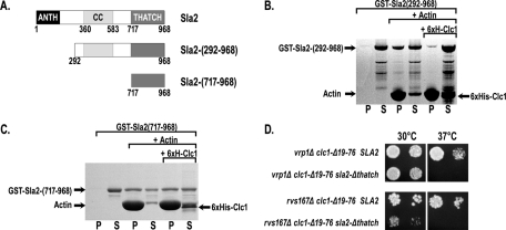
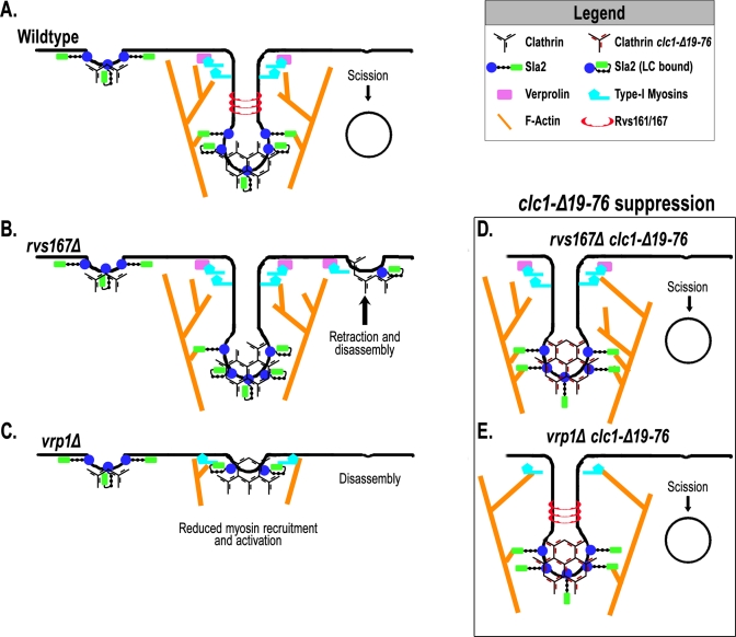
The role of clathrin light chain (CLC) in clathrin-mediated endocytosis is not completely understood. Previous studies showed that the CLC N-terminus (CLC-NT) binds the Hip1/Hip1R/Sla2 family of membrane/actin-binding factors and that overexpression of the CLC-NT in yeast suppresses endocytic defects of clathrin heavy-chain mutants. To elucidate the mechanistic basis for this suppression, we performed synthetic genetic array analysis with a clathrin CLC-NT deletion mutation (clc1-Δ19-76). clc1-Δ19-76 suppressed the internalization defects of null mutations in three late endocytic factors: amphiphysins (rvs161 and rvs167) and verprolin (vrp1). In actin sedimentation assays, CLC binding to Sla2 inhibited Sla2 interaction with F-actin. Furthermore, clc1-Δ19-76 suppression of the rvs and vrp phenotypes required the Sla2 actin-binding talin-Hip1/R/Sla2 actin-tethering C-terminal homology domain, suggesting that clc1-Δ19-76 promotes internalization by prolonging actin engagement by Sla2. We propose that CLC directs endocytic progression by pruning the Sla2-actin attachments in the clathrin lattice, providing direction for membrane internalization.


Figures

FIGURE 1:
FIGURE 1:
Synthetic genetic array analysis with the clc1-Δ19-76 allele. (A) Schematics of Clc1 and Clc1-Δ19-76, highlighting regions that bind Sla2 (blue), CHC (gray), or calmodulin (yellow) and the region comprising an EF hand motif (green). (B) Immunoblot of protein extracts from wild type (SL1462), clc1-Δ19-76 (SL5677), and clc1Δ (SL1620) probed with anti-Clc1 and anti-Pgk1 (loading control). (C) Same as B, but probed with anti-Chc1 monoclonal antibodies. (D–G) Edges represent published physical protein–protein interactions; nodes are white if they were not in the screen and gray if no interaction was identified. If double mutants produced a synthetic growth defect relationship, they are labeled in red, and nodes demonstrating synthetic rescue with clc1-Δ19-76 are labeled in green. (D) clc1-Δ19-76 demonstrated a relationship with three AP-1–complex subunits (p ≤ 0.01). (E) Four of the five core retromer complex proteins were identified (p ≤ 0.01). (F) Three of the five components of the GET complex were identified (p ≤ 0.01). (G) Endocytic network showing hits from SGA analysis with clc1-Δ19-76.
FIGURE 2:
FIGURE 2:
clc1-Δ19-76 suppresses the growth, actin polarity, and fluid-phase endocytosis defects of verprolin and amphiphysin null mutants. (A) Growth with vrp1Δ: wild type (SL1462), clc1-Δ19-76 (SL6044), vrp1Δ (SL4136), and vrp1Δ clc1-Δ19-76 (SL6049) were fivefold serially diluted, plated on YEPD, and grown at 30 or 37°C for 60 h. (B) Growth with rvs167Δ: wild type (SL1462), clc1-Δ19-76 (SL6044), rvs167Δ (RH2951), and rvs167Δ clc1-Δ19-76 (SL6052) were plated and grown as in A. (C) Actin polarization: strains shown in A and B were grown at 25°C, fixed, and stained with Alexa Fluor 568–phalloidin. Each image is a max-Z projection of 12 (0.2 μm) optical sections after nearest-neighbor deconvolution (bar, 5 μm). (D) Quantification of actin polarization reported as percentage of small and medium budded cells with >50% of phalloidin stain in the bud (n = 75). (E) Example micrographs of strains indicated in A and B after Lucifer yellow (LY) uptake for 1 h at 25°C (bar, 5 μm). (F) Quantification of LY uptake reported as percentage of cells with vacuolar LY (n = 90).
FIGURE 3:
FIGURE 3:
Defects in endocytic dynamics of vrp1Δ are suppressed when combined with clc1-Δ19-76. (A) Representative kymographs of Sla1-GFP/Abp1-RFP patches in wild type (SL5311), clc1-Δ19-76 (SL6068), vrp1Δ (SL6065), and vrp1Δ clc1-Δ19-76 (SL6062). (B) Representative kymographs of Sla2-GFP/Abp1-RFP patches in wild type (SL5927), clc1-Δ19-76 (SL6084), vrp1Δ (SL6081), vrp1Δ clc1-Δ19-76 (SL6077). (C) Fluorescence lifetimes of Sla2-GFP, Sla1-GFP, and Abp1-RFP in strains shown in A and B. Data are reported as average ± SD (n ≥ 50). †p ≤ 0.0001 vs. wild type; ‡p ≤ 0.0001 vs. vrp1Δ; #p ≤ 0.001 vs. vrp1Δ. (D) Tangential kymographs illustrating inward movement of Sla2-GFP/Abp1-RFP endocytic patches in wild type, vrp1Δ, and vrp1Δ clc1-Δ19-76. (E) Percentage of Sla2-GFP/Abp1-RFP patches that demonstrate “normal” inward movement (n = 90). (F) Example plots of Sla2-GFP trajectories comparing inward movement from wild type, vrp1Δ, and vrp1Δ clc1-Δ19-76. Each point represents a 2-s frame, with the length of the lines between frames indicating distance moved. Initial coordinates are highlighted in green, and the final time points are in red.
FIGURE 4:
FIGURE 4:
Defects in endocytic dynamics of rvs167Δ are suppressed when combined with clc1-Δ19-76. (A) Representative kymographs of Sla1-GFP/Abp1-RFP patches in wild type (SL5311), clc1-Δ19-76 (SL6068), rvs167Δ (SL6108), and rvs167Δ clc1-Δ19-76 (SL6191). (B) Representative kymographs of Sla2-GFP/Abp1-RFP patches in wild type (SL5927), clc1-Δ19-76 (SL6084), rvs167Δ (SL6196), and rvs167Δ clc1-Δ19-76 (SL6197). (C) Fluorescence lifetimes of Sla2-GFP, Sla1-GFP, and Abp1-RFP in strains shown in A and B. Data are reported as average ± SD (n ≥ 50). †p ≤ 0.0001 vs. wild type; ‡p ≤ 0.0001 vs. rvs167Δ; #p ≤ 0.03 vs. rvs167Δ. (D) Percentage of patches that show “normal” inward movement following Abp1 accumulation (n = 90). Similar results were obtained for rvs161Δ (see Supplemental Figure S1).
FIGURE 5:
FIGURE 5:
vrp1Δ and rvs167Δ are not suppressed by a clathrin LC null (clc1Δ). (A) Wild type (SL1462), clc1Δ (SL1620), vrp1Δ (SL4136), and vrp1Δ clc1Δ (SL6290) were fivefold serially diluted, plated on YEPD, and grown at 30 or 37°C for 60 h. (B) Kymographs of Sla2-GFP and Abp1-RFP in vrp1Δ clc1Δ (SL6291) illustrating the types of dynamic behaviors observed and percentages of each (n = 60). (C) Wild type (SL1462), clc1Δ (SL1620), rvs167Δ (RH2951), and rvs167Δ clc1Δ (SL6292) were plated and grown as indicated in A. (D) Kymographs of Sla2-GFP and Abp1-RFP in rvs167Δ clc1Δ (SL6293) illustrating the types of dynamic behaviors and percentages of each (n = 60).
FIGURE 6:
FIGURE 6:
Clathrin LC inhibits Sla2 binding to F-actin. (A) GST-Sla2 fusions used in actin binding assays. Schematic highlights sequences corresponding to the N-terminal ANTH domain (black), coiled-coil domain (light gray), and THATCH domain (dark gray). (B) Actin sedimentation assays performed using 3 μM GST-Sla2-292-968, with or without 10 μM F-actin and with or without 15 μM 6xHis-Clc1. (C) Actin sedimentation assays performed as in B using 3 μM GST-Sla2-717-968.D. Sla2 actin-binding THATCH domain is necessary for clc1-Δ19-76 suppression: strains were fivefold serially diluted, plated on YEPD, and grown at 30 and 37°C. Strains are vrp1Δ clc1-Δ19-76 (SL6230), vrp1Δ clc1-Δ19-76 sla2-Δthatch (SL6231), rvs167Δ clc1-Δ19-76 (SL6238), and rvs167Δ clc1-Δ19-76 sla2-Δthatch (SL6239).
FIGURE 7:
FIGURE 7:
Endocytic defects caused by elimination of the type I myosins (myo3Δ myo5Δ) or WASp (las17Δ) are suppressed by clc1-Δ19-76. (A) Growth with myo3Δ myo5Δ: wild type (SL1462), clc1-Δ19-76 (SL6044), myo3Δ myo5Δ (SL6561), myo3Δ myo5Δ clc1-Δ19-76 (SL6576), myo5Δ (SL6580), and myo5Δ clc1-Δ19-76 (SL6579) were fivefold serially diluted, plated on YEPD, and grown at 30 or 37°C for 60 h. (B) Growth of las17Δ (SL6602) and las17Δ clc1-Δ19-76 (SL6603) as in A. (C) Representative kymographs of Sla1-GFP/Abp1-RFP patches in wild type (SL5311), myo5Δ (SL6579), myo5Δ clc1-Δ19-76 (SL6554), myo3Δ myo5Δ (SL6562), and myo3Δ myo5Δ clc1-Δ19-76 (SL6575). (D) Representative kymographs of Sla2-GFP/Abp1-RFP patches in wild type (SL5311), myo5Δ (SL6569), myo5Δ clc1-Δ19-76 (SL6572), myo5Δ myo3Δ (SL6566), and myo3Δ myo5Δ clc1-Δ19-76 (SL6568). (E) Representative kymographs of Sla1-GFP/Abp1-RFP patches in las17Δ (SL6596) and las17Δ clc1-Δ19-76 (SL6597). (F) Representative kymographs of Sla2-GFP/Abp1-RFP patches in las17Δ (SL6598) and las17Δ clc1-Δ19-76 (SL6600). (G) Fluorescence lifetimes of Sla2-GFP, Sla1-GFP, and Abp1-RFP in strains shown in C–F. Data are reported as average ± SD (n ≥ 50). †p ≤ 0.0009 vs. wild type; *p ≤ 0.004 vs. wild type; #p ≤ 0.0001 vs. myo3Δ myo5Δ; °p ≤ 0.0003 vs. myo5Δ; ‡p ≤ 0.0001 vs. las17Δ.
FIGURE 8:
FIGURE 8:
Model for CLC regulation of Sla2 binding to F-actin at endocytic patches. (A–C) Model of endocytic patches in wild type (A), rvs167Δ (B), and vrp1Δ (C). (D, E) Models for how preventing CLC regulation of the Sla2–actin interaction helps to overcome amphiphysin (D) and verprolin (E) mutants by increasing attachments to F-actin. Suppression of myo3/5Δ and las17Δ is similar to that of vrp1Δ.

References

    1. Anderson BL, Boldogh I, Evangelista M, Boone C, Greene LA, Pon LA. The Src homology domain 3 (SH3) of a yeast type I myosin, Myo5p, binds to verprolin and is required for targeting to sites of actin polarization. J Cell Biol. 1998;141:1357–1370. - PMC - PubMed
    1. Bader GD, Hogue CW. An automated method for finding molecular complexes in large protein interaction networks. BMC Bioinformatics. 2003;4:2. - PMC - PubMed
    1. Barker SL, Lee L, Pierce BD, Maldonado-Baez L, Drubin DG, Wendland B. Interaction of the endocytic scaffold protein Pan1 with the type I myosins contributes to the late stages of endocytosis. Mol Biol Cell. 2007;18:2893–2903. - PMC - PubMed
    1. Bensen ES, Costaguta G, Payne GS. Synthetic genetic interactions with temperature-sensitive clathrin in Saccharomyces cerevisiae. Roles for synaptojanin-like Inp53p and dynamin-related Vps1p in clathrin-dependent protein sorting at the trans-Golgi network. Genetics. 2000;154:83–97. - PMC - PubMed
    1. Bensen ES, Yeung BG, Payne GS. Ric1p and the Ypt6p GTPase function in a common pathway required for localization of trans-Golgi network membrane proteins. Mol Biol Cell. 2001;12:13–26. - PMC - PubMed

Publication types

MeSH terms