. 2011 Oct;22(19):3734-46.
doi: 10.1091/mbc.E10-11-0917. Epub 2011 Aug 17.

The activation of ezrin-radixin-moesin proteins is regulated by netrin-1 through Src kinase and RhoA/Rho kinase activities and mediates netrin-1-induced axon outgrowth

Affiliations

The activation of ezrin-radixin-moesin proteins is regulated by netrin-1 through Src kinase and RhoA/Rho kinase activities and mediates netrin-1-induced axon outgrowth

Judith Antoine-Bertrand et al. Mol Biol Cell. 2011 Oct.

Abstract

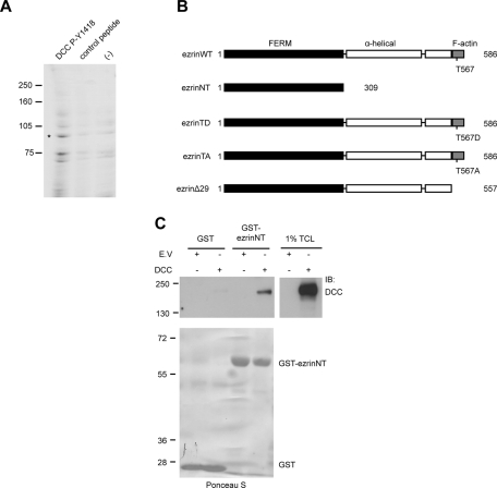
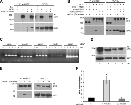
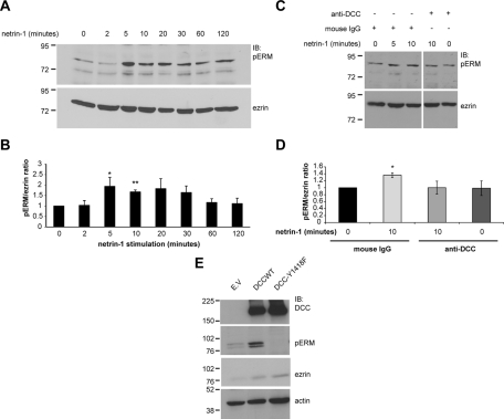
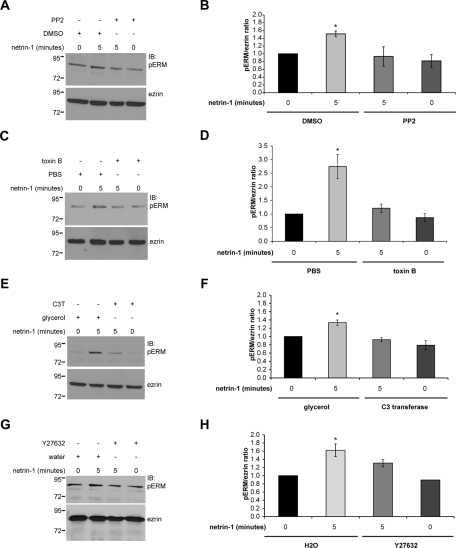
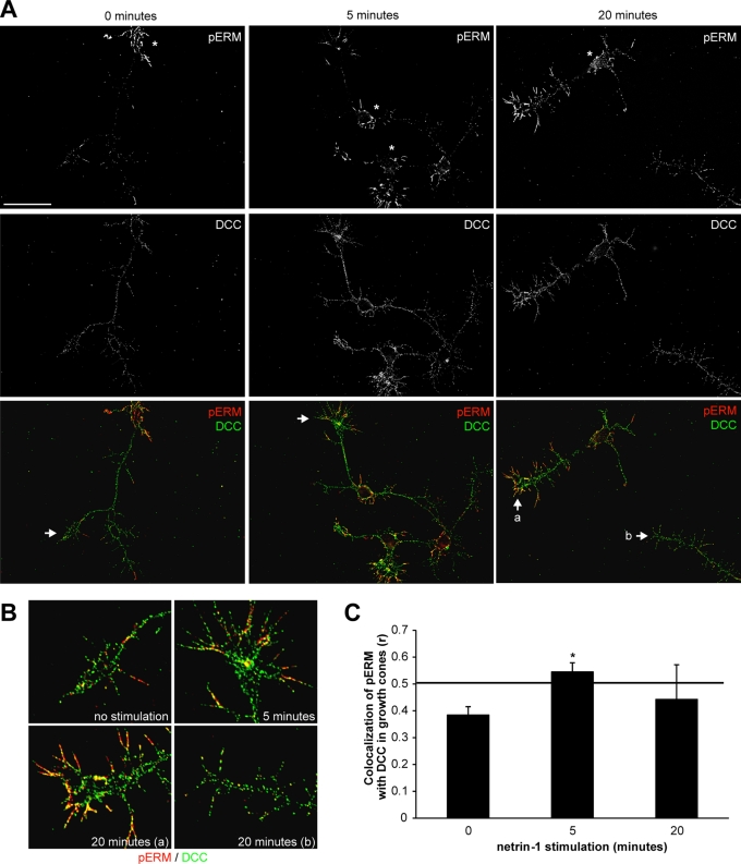
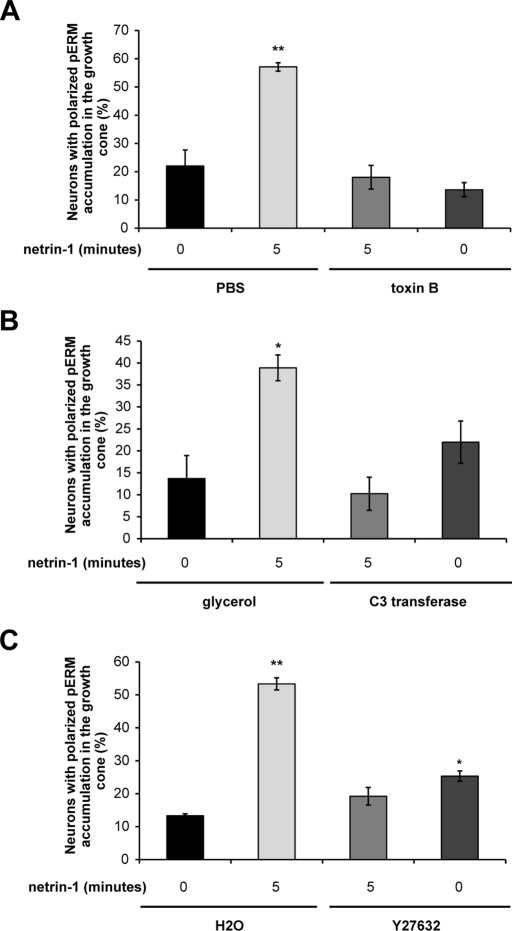
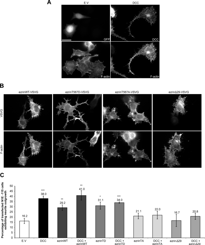
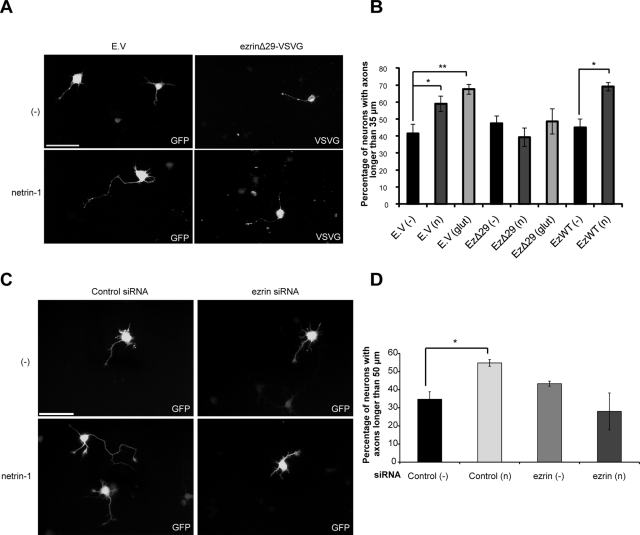
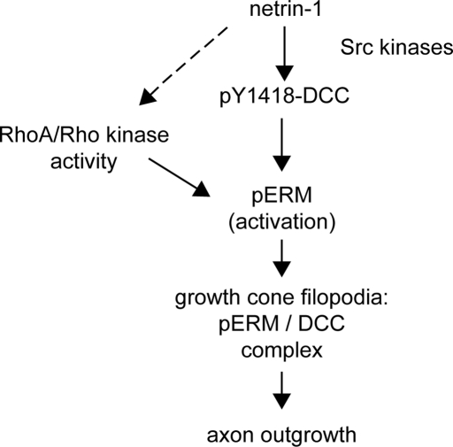
The receptor Deleted in Colorectal Cancer (DCC) mediates the attractive response of axons to the guidance cue netrin-1 during development. On netrin-1 stimulation, DCC is phosphorylated and induces the assembly of signaling complexes within the growth cone, leading to activation of cytoskeleton regulators, namely the GTPases Rac1 and Cdc42. The molecular mechanisms that link netrin-1/DCC to the actin machinery remain unclear. In this study we seek to demonstrate that the actin-binding proteins ezrin-radixin-moesin (ERM) are effectors of netrin-1/DCC signaling in embryonic cortical neurons. We show that ezrin associates with DCC in a netrin-1-dependent manner. We demonstrate that netrin-1/DCC induces ERM phosphorylation and activation and that the phosphorylation of DCC is required in that context. Moreover, Src kinases and RhoA/Rho kinase activities mediate netrin-1-induced ERM phosphorylation in neurons. We also observed that phosphorylated ERM proteins accumulate in growth cone filopodia, where they colocalize with DCC upon netrin-1 stimulation. Finally, we show that loss of ezrin expression in cortical neurons significantly decreases axon outgrowth induced by netrin-1. Together, our findings demonstrate that netrin-1 induces the formation of an activated ERM/DCC complex in growth cone filopodia, which is required for netrin-1-dependent cortical axon outgrowth.


Figures

FIGURE 1:
FIGURE 1:
Ezrin interacts with DCC. (A) E13 rat brain protein lysates were incubated with Affi-Gel-DCC-P-Y1418 phosphopeptide, Affi-Gel-DCC unphosphorylated peptide (control), or Affi-Gel beads (−). Bound proteins were resolved by SDS–PAGE and stained with Coomassie blue. Peptides matching the ERM protein sequences of ezrin, radixin, and moesin were identified by MS/MS analysis of the band at approximately 85 kDa represented by the asterisk. (B) The domain architecture of wild-type ezrin (ezrinWT) and ezrin mutant proteins. EzrinWT consists of the N-terminal FERM domain, the α-helical linker region, and the C-terminal F-actin–binding domain. (C) Lysates of HEK293 cells transfected with pRK5 or pRK5-DCC were incubated with GST or GST-ezrinNT fusion proteins coupled to glutathione–agarose beads (n = 4). GST pull-down–associated proteins and 1% of total cell lysates (TCL) were resolved by SDS–PAGE and immunoblotted (IB) with antibodies against DCC. Purified GST proteins were stained with Ponceau S prior to immunoblotting (bottom).
FIGURE 2:
FIGURE 2:
Netrin-1 regulates the interaction between ezrin and DCC. Protein lysates of N1E-115 cells (A) and HEK293 cells (B) transfected with empty vector (E.V.), pRK5-DCC, pRK5-DCC (1–1120), VSVG-tagged ezrinTD, ezrin wild type, or ezrinΔ29 as indicated or (E) lysates of embryonic rat cortical neurons (E18, 2DIV) stimulated with netrin-1 for various periods of time were submitted to immunoprecipitation (IP) using anti-DCC antibodies. Immunoprecipitated proteins and 1% (A, B) or 10% (E) of total cell lysates (TCL) were resolved by SDS–PAGE, followed by immunoblotting (IB) using anti-DCC, anti-ezrin, or anti-VSVG antibodies. mRNA (C) or proteins (D) were extracted from E18 cortices (0DIV) or cultured cortical neurons (1, 2, and 3 DIV). (C) RT-PCR amplification of cDNA from ezrin (233 base pairs), moesin (205 base pairs), radixin (242 base pairs), merlin (248 base pairs), and GAPDH (207 base pairs). Control reactions (c) were performed with water instead of cDNA for each primer pair. (D) Protein lysates of cortical neurons were resolved by SDS–PAGE and immunoblotted with antibodies against DCC or ezrin. (F) Quantitative densitometry of the amount of ezrin immunoprecipitated with DCC in E represented as the fold increase relative to unstimulated neurons (n = 3). The increase in the amount of ezrin immunoprecipitated is significant after 5 min of netrin-1 stimulation (*p < 0.01).
FIGURE 3:
FIGURE 3:
Netrin-1 induces the phosphorylation of ERM proteins through DCC in embryonic cortical neurons. (A) Protein lysates of embryonic rat cortical neurons (E18, 2DIV) stimulated with netrin-1 for various periods of time or (E) protein lysates of NIE-115 cells transfected with empty vector (E.V.), pRK5-DCC, or pRK5-DCC-Y1418F were resolved by SDS–PAGE, followed by immunoblotting using anti-DCC, anti-pERM, anti-ezrin, or anti-actin antibodies. (B) Quantitative densitometry of A is represented as the ratio of pERM over total ezrin and corresponds to the average of at least three independent experiments. The increase in pERM levels after 5 and 10 min of netrin-1 stimulation was significant (*p < 0.05; **p < 0.005). (C) The experiment was as described in A, except that function-blocking DCC antibodies were added before netrin-1 stimulation. Mouse IgGs were used as a negative control. (D) Quantitative densitometry of C is represented as the ratio of pERM over total ezrin and corresponds to the average of at least three independent experiments. The increase in pERM after 10 min of netrin-1 stimulation is significant (*p < 0.05). Error bars, SEM. (E) The upper pERM band represents ezrin and radixin, and the lower band is moesin.
FIGURE 4:
FIGURE 4:
Src family kinases and RhoA/Rho kinase mediate ERM protein phosphorylation in cortical neurons stimulated with netrin-1. Embryonic rat cortical neurons (E18, 2DIV) were incubated with netrin-1 for the indicated times following treatment with (A, B) Src kinase inhibitor PP2 or DMSO as a negative control, (C, D) Rho GTPase inhibitor toxin B or PBS as a negative control, (E, F) RhoA inhibitor C3 transferase (C3T) or glycerol as a negative control, or (G, H) Rho kinase inhibitor Y27632 or water (H2O) as a negative control. Protein lysates were resolved by SDS–PAGE and immunoblotted with anti-pERM or anti-ezrin antibodies. Quantitative densitometry is represented as the ratio of pERM over total ezrin and corresponds to the average of at least three independent experiments. The increase in pERM after 5 min of netrin-1 stimulation was significant (*p < 0.05), whereas there was no significant increase after the PP2, toxin B, C3 transferase, or Y27632 treatment compared with unstimulated neurons (0 min). Error bars, SEM.
FIGURE 5:
FIGURE 5:
Netrin-1 induces the accumulation of phosphorylated ERM proteins and their colocalization with DCC in growth cone filopodia. (A) Embryonic rat cortical neurons (E18, 2DIV) were incubated with netrin-1 for the indicated times, fixed, and immunostained with antibodies against DCC (green) and pERM (red). Cell bodies are represented by asterisks. Scale bar, 5 μm. (B) Magnification of regions designated by arrows in A. Growth cones after 20 min of netrin-1 stimulation (a) with pERM-DCC colocalization, and (b) with no significant pERM-DCC colocalization. (C) Quantification of pERM–DCC colocalization in growth cones using Pearson's correlation coefficient r; *p < 0.05. The minimal r value for significant pERM-DCC colocalization is 0.5 (horizontal line). Error bars, SEM.
FIGURE 6:
FIGURE 6:
RhoA/Rho kinase is required to mediate the accumulation of phosphorylated ERM proteins in growth cones upon netrin-1 stimulation. Embryonic rat cortical neurons (E18, 2DIV) were incubated with netrin-1 for the indicated times following treatment with (A) Rho GTPase inhibitor toxin B or PBS as a negative control, (B) RhoA inhibitor C3 transferase (C3T) or glycerol as a negative control, or (C) Rho kinase inhibitor Y27632 or water (H2O) as a negative control. Neurons were fixed and immunostained with anti-pERM and anti-DCC antibodies (Supplemental Figure S3). The quantification of the accumulation of phosphorylated ERM proteins (pERM) in growth cones corresponds to three independent experiments. Student's unpaired t test was used for statistical analysis, and the data are presented as mean percentage ± SEM (*p < 0.05; **p < 0.005).
FIGURE 7:
FIGURE 7:
Impaired ezrin activity inhibits DCC-mediated neurite outgrowth. (A, B) N1E-115 cells were transfected with either pEGFP (E.V.), pRK5-DCC, pCB6-ezrin-VSVG (ezrinWT), pCB6-ezrinT567D-VSVG (ezrinTD), pCB6-ezrinT567A-VSVG (ezrinTA), or pCB6-ezrinΔ29-VSVG (ezrinΔ29), fixed, and immunostained with antibodies against DCC or VSVG. F-Actin was labeled with phalloidin-TRITC. Scale bar, 5 μm. (C) The percentage of transfected N1E-115 cells with neurites longer than their cell body was measured 24 h posttransfection. The values correspond to the average of at least three independent experiments in which at least 150 transfected cells were counted. Error bars, SEM (*p < 0.05; **p < 0.01; ***p < 0.001).
FIGURE 8:
FIGURE 8:
Dominant-negative ezrin and ezrin down-regulation inhibit netrin-1–mediated axon outgrowth. (A, B) Embryonic rat cortical neurons (E18) were transfected at 1DIV with pEGFP (empty vector, E.V.), pCB6-ezrin-VSVG (ezWT), or pCB6-ezrinΔ29-VSVG (ezΔ29) and incubated for 24 h with netrin-1 or control medium. Neurons were fixed at 2DIV and immunostained with anti-VSVG antibodies. (C, D) Embryonic rat cortical neurons (E18) were electroporated at 0DIV with pmaxGFP and Negative Control siRNA or with pmaxGFP and ezrin siRNA to down-regulate ezrin (Supplemental Figure S2, B and C) and incubated for 24 h with netrin-1 or control medium. Quantification of axon outgrowth in transfected cells following a 24-h incubation with control medium (−), netrin-1 (n), or glutamate (glut) is represented as the percentage of transfected cortical neurons with axons longer than 35 μm (B) or 50 μm (D) in length. The values correspond to the average of at least three independent experiments. Error bars, SEM (*p < 0.05; **p < 0.001). Scale bar, 50 μm.
FIGURE 9:
FIGURE 9:
Regulation and function of the ERM–DCC complex downstream of netrin-1 in cortical neurons. Netrin-1 promotes tyrosine phosphorylation of DCC via Src kinases. The signaling cascade downstream of netrin-1 and its phosphorylated receptor promotes the phosphorylation of ERM protein in concert with RhoA and its effector, Rho kinase. The mechanism by which netrin-1 activates RhoA has yet to be determined (dashed arrow). Upon phosphorylation, activated ERM proteins (pERM) accumulate in growth cone filopodia, where they form a complex with DCC. The phosphorylation of the receptor on Y1418 positively regulates ERM protein activation, thus promoting ERM–DCC complex assembly. Ultimately, the activity of ERM proteins promotes axon outgrowth downstream of netrin-1, likely through their interaction with both DCC and the actin cytoskeleton in growth cone filopodia.

References

    1. Algrain M, Turunen O, Vaheri A, Louvard D, Arpin M. Ezrin contains cytoskeleton and membrane binding domains accounting for its proposed role as a membrane-cytoskeletal linker. J Cell Biol. 1993;120:129–139. - PMC - PubMed
    1. Barret C, Roy C, Montcourrier P, Mangeat P, Niggli V. Mutagenesis of the phosphatidylinositol 4,5-bisphosphate (PIP(2)) binding site in the NH(2)-terminal domain of ezrin correlates with its altered cellular distribution. J Cell Biol. 2000;151:1067–1080. - PMC - PubMed
    1. Berryman M, Gary R, Bretscher A. Ezrin oligomers are major cytoskeletal components of placental microvilli: a proposal for their involvement in cortical morphogenesis. J Cell Biol. 1995;131:1231–1242. - PMC - PubMed
    1. Briancon-Marjollet A, et al. Trio mediates netrin-1-induced Rac1 activation in axon outgrowth and guidance. Mol Cell Biol. 2008;28:2314–2323. - PMC - PubMed
    1. Charrin S, Alcover A. Role of ERM (ezrin-radixin-moesin) proteins in T lymphocyte polarization, immune synapse formation and in T cell receptor-mediated signaling. Front Biosci. 2006;11:1987–1997. - PubMed

Publication types

MeSH terms