. 2011 Oct 14;286(41):35955-35965.
doi: 10.1074/jbc.M111.276238. Epub 2011 Aug 17.

Orphan macrodomain protein (human C6orf130) is an O-acyl-ADP-ribose deacylase: solution structure and catalytic properties

Affiliations

Orphan macrodomain protein (human C6orf130) is an O-acyl-ADP-ribose deacylase: solution structure and catalytic properties

Francis C Peterson et al. J Biol Chem. .

Abstract

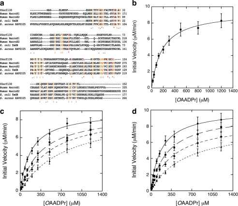
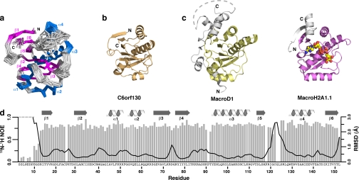
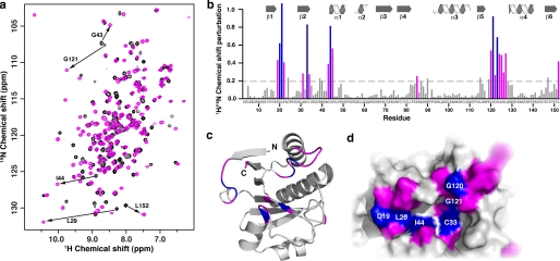
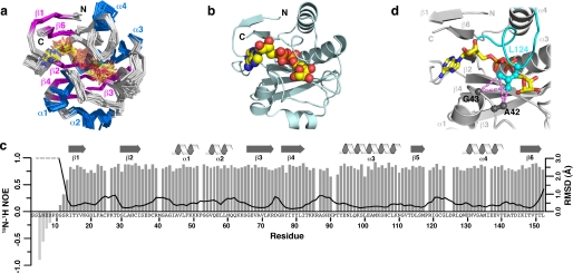
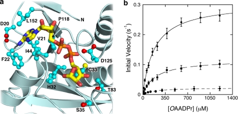
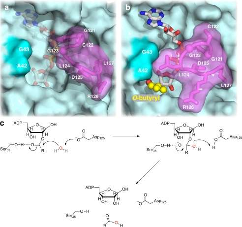
Post-translational modification of proteins/histones by lysine acylation has profound effects on the physiological function of modified proteins. Deacylation by NAD(+)-dependent sirtuin reactions yields as a product O-acyl-ADP-ribose, which has been implicated as a signaling molecule in modulating cellular processes. Macrodomain-containing proteins are reported to bind NAD(+)-derived metabolites. Here, we describe the structure and function of an orphan macrodomain protein, human C6orf130. This unique 17-kDa protein is a stand-alone macrodomain protein that occupies a distinct branch in the phylogenic tree. We demonstrate that C6orf130 catalyzes the efficient deacylation of O-acetyl-ADP-ribose, O-propionyl-ADP-ribose, and O-butyryl-ADP-ribose to produce ADP-ribose (ADPr) and acetate, propionate, and butyrate, respectively. Using NMR spectroscopy, we solved the structure of C6orf130 in the presence and absence of ADPr. The structures showed a canonical fold with a deep ligand (ADPr)-binding cleft. Structural comparisons of apo-C6orf130 and the ADPr-C6orf130 complex revealed fluctuations of the β(5)-α(4) loop that covers the bound ADPr, suggesting that the β(5)-α(4) loop functions as a gate to sequester substrate and offer flexibility to accommodate alternative substrates. The ADPr-C6orf130 complex identified amino acid residues involved in substrate binding and suggested residues that function in catalysis. Site-specific mutagenesis and steady-state kinetic analyses revealed two critical catalytic residues, Ser-35 and Asp-125. We propose a catalytic mechanism for deacylation of O-acyl-ADP-ribose by C6orf130 and discuss the biological implications in the context of reversible protein acylation at lysine residues.


Figures

FIGURE 1.
FIGURE 1.
C6orf130 deacetylates OAADPr. a, sequence alignment C6orf130 with other MacroD-like proteins human MacroD1, human MacroD2, E. coli YmdB, and S. aureus SAV0325. b, steady-state kinetic analysis of C6orf130. The deacetylation reaction of OAADPr catalyzed by C6orf130 follows saturation kinetics. The steady-state kinetic parameters are determined by radioactive assays for acetate formation and HPLC assays for ADP-ribose formation. Apparent Km as measured was 182 ± 17 μm; the kcat as measured was 0.31 ± 0.03 s−1. The reaction mixtures contained 0.5 μm C6orf130. c, inhibition of C6orf130 deacetylase activity by ADPr. The deacetylation reactions were carried out in 50 mm Tris-HCl, pH 7.3, containing 0.5 μm C6orf130. The reactions were initiated in the presence of 0 μm (circles), 100 μm (squares), 200 μm (diamonds), and 400 μm (triangles) ADPr, respectively. The Km′ values measured were 183.3, 340.6, 480.1, and 782.2 μm in the presence of 0, 100, 200, and 400 μm initial ADPr, respectively. The Vmax values were 0.31, 0.29, 0.29, and 0.30 s−1. The Ki was determined to be 119.3 ± 4.5 μm. The error bars represent standard deviations calculated from the measured initial velocities at each substrate concentration from three separate experiments. d, inhibition of C6orf130 deacetylase activity by NAADPr. The deacetylation reactions were carried out under the same conditions as described in c in the presence of 0 μm (circles), 125 μm (squares), 250 μm (diamonds), and 500 μm (triangles) NAADPr, respectively. The Km′ values measured were 183.3, 340.6, 480.1, and 782.2 μm in the presence of 0, 125, 250, and 500 μm initial NAADPr, respectively. The Vmax values were 0.31, 0.29, 0.29, and 0.30 s−1. The Ki was determined to be 223.3 ± 11.4 μm. The error bars represent standard deviations calculated from the initial velocities at each substrate concentration from three separate experiments.
FIGURE 2.
FIGURE 2.
NMR structure of the C6orf130 macrodomain. a, ensemble of the final 20 NMR structures (Cα trace). α-Helices and β-sheets are shown in blue and magenta, respectively. Residues 3–12 are unstructured and have been omitted for clarity. b, ribbon view of C6orf130 showing the core α-β-α sandwich fold typical of macrodomains. c, the MacroD1 (yellow; PDB code 2X47) and MacroH2A1.1 (magenta; PDB code 3IID) macrodomains contain the central α-β-α sandwich but also contain extended structural features at the N and C termini (gray). d, 15N-1H heteronuclear NOE values and global backbone atomic r.m.s.d. values plotted as a function of residue number. Values are missing for Gly-123 and Leu-124. Global r.m.s.d. values were calculated with the ensemble superimposed using residues 14–120 and 126–152.
FIGURE 3.
FIGURE 3.
C6orf130 binds ADPr specifically. a, 15N-1H HSQC of C6orf130 in the apo (black) and ADPr bound states (magenta). b, combined 1H/15N chemical shift perturbations in response to ADPr binding were calculated and plotted as a function of the C6orf130 residue number. Residues with chemical shift perturbations of ≥0.25 were mapped onto the C6orf130 apostructure and are shown in ribbon (c) and surface (d) views. The perturbations localize to one face of the molecule and identify the conserved binding cleft as the ADPr-binding site. Chemical shift perturbations ranging from 0.25 to 0.59 and those ≥0.6 are shown in magenta and blue, respectively.
FIGURE 4.
FIGURE 4.
NMR structure of the C6orf130-ADPr complex. a, ensemble of the final 20 NMR structures (Cα trace). α-Helices and β-sheets are shown in blue and magenta, respectively. Residues 3–12 are unstructured and have been omitted for clarity. b, ribbon view of C6orf130-ADPr complex with ADPr shown in yellow. c, 15N-1H heteronuclear NOE values and global backbone atomic r.m.s.d. values plotted as a function of residue number. 15N-1H heteronuclear NOE values are missing or excluded for residues Met-40, Phe-52, Arg-86, Leu-124, Asp-125, and Ile-136. Global r.m.s.d. values were calculated with the ensemble superimposed using residues 14–152. d, NOEs between the methyl groups of Leu-124 in the β54 loop (cyan) and residues Ala-42 and Gly-43 (dark gray) are present only in the ADPr-C6orf130 complex. NOEs are shown in magenta as dashed lines.
FIGURE 5.
FIGURE 5.
Kinetic comparison of wild type C6orf130 and its variants. a, key amino acid residues interacting with the bound ADPr. Side chains are shown in ball-and-stick form, and oxygen atoms are highlighted in red. b, Thr-83 and Ser-35 located in the vicinity of the 2- and 3-hydroxyl groups of the bound ADPr in the MacroD1 are mutated to alanine. A steady-state kinetic comparison of the wild type C6orf130 (circles) and the T83A (squares) and S35A (diamonds) mutants shows that these residues are important to catalysis. Substituting Thr-83 and Ser-35 with Ala mainly affects the Vmax. The Km values are only minimally affected. The error bars represent standard deviations calculated from the measured initial velocities at each substrate concentration from three separate experiments. Data points of mutant partial activities are the mean values from three separate measurements.
FIGURE 6.
FIGURE 6.
Substrate binding and catalytic mechanism of C6orf130. a and b, C6orf130 accommodates longer acyl chains. A model of OBADPr bound in the active site of C6orf130 was constructed by addition of the butyryl moiety (yellow ball and stick) to the 2-hydroxyl of the distal ribose. a, in apo-C6orf130, residues 120–127 of the β54 loop (magenta) are in the open position, exposing the ligand-binding cleft (indicated by ADPr). b, ADPr binding dampens the fluctuations of the β54 loop (magenta) closing the gate that sequesters ADPr or other ligands in the C6orf130 active site. Lid closure forms a pliable pore through which longer acyl chains, such as the butyryl moiety modeled here (yellow ball and stick), can be accommodated in the C6orf130 active site. c, proposed catalytic mechanism of C6orf130. This mechanism is based on structural analyses of both the apo-C6orf130 and the C6orf130-ADPr complex, mutagenesis of key residues in the vicinity of the distal ribose of the bound ADPr, and kinetic studies of the mutant enzymes.

References

    1. Anantharaman V., Aravind L. (2002) Genome Biol. 3, RESEARCH0061 - PMC - PubMed
    1. Pehrson J. R., Fuji R. N. (1998) Nucleic acids Res. 26, 2837–2842 - PMC - PubMed
    1. Allen M. D., Buckle A. M., Cordell S. C., Löwe J., Bycroft M. (2003) J. Mol. Biol. 330, 503–511 - PubMed
    1. Ladurner A. G. (2003) Mol. Cell 12, 1–3 - PubMed
    1. Till S., Ladurner A. G. (2009) Front. Biosci. 14, 3246–3258 - PubMed

Publication types