Comparative Study
. 2011 Aug 17;31(33):11756-61.
doi: 10.1523/JNEUROSCI.1960-11.2011.

APP is phosphorylated by TrkA and regulates NGF/TrkA signaling

Affiliations
Comparative Study

APP is phosphorylated by TrkA and regulates NGF/TrkA signaling

Carmela Matrone et al. J Neurosci. .

Abstract

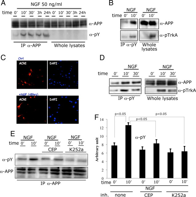
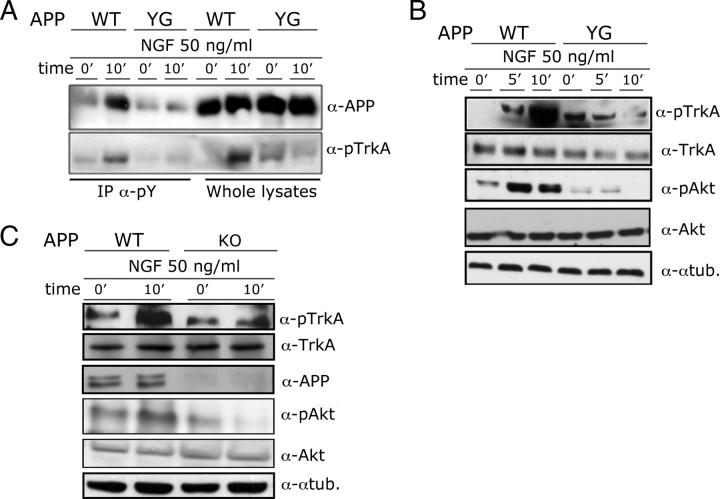
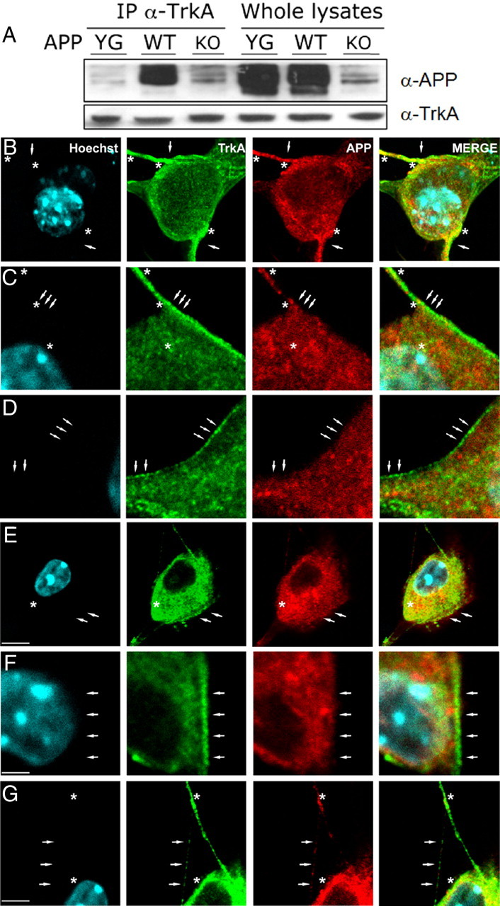
The pathogenic model of Alzheimer's disease (AD) posits that aggregates of amyloid β, a product of amyloid precursor protein (APP) processing, cause dementia. However, alterations of normal APP functions could contribute to AD pathogenesis, and it is therefore important to understand the role of APP. APP is a member of a gene family that shows functional redundancy as documented by the evidence that single knock-out mice are viable, whereas mice with combined deletions of APP family genes die shortly after birth. A residue in the APP intracellular region, Y(682), is indispensable for these essential functions of APP. It is therefore important to identify pathways that regulate phosphorylation of Y(682) as well as the role of Y(682) in vivo. TrkA is associated with both phosphorylation of APP-Y(682) and alteration of APP processing, suggesting that tyrosine phosphorylation of APP links APP processing and neurotrophic signaling to intracellular pathways associated with cellular differentiation and survival. Here we have tested whether the NGF/TrkA signaling pathway is a physiological regulator of APP phosphorylation. We find that NGF induces tyrosine phosphorylation of APP, and that APP interacts with TrkA and this interaction requires Y(682). Unpredictably, we also uncover that APP, and specifically Y(682), regulates activation of the NGF/TrkA signaling pathway in vivo, the subcellular distribution of TrkA and the sensitivity of neurons to the trophic action of NGF. This evidence suggests that these two membrane protein's functions are strictly interconnected and that the NGF/TrkA signaling pathway is involved in AD pathogenesis and can be used as a therapeutic target.


Figures

Figure 1.
Figure 1.
NGF triggers phosphorylation of APP via the TrkA receptor. A, Primary cortical neurons from WT mice were treated with 50 ng/ml NGF for the indicated time periods. Lysates from treated neurons were immunoprecipitated with an α-APP antibody (IP-α-APP). Immunoprecipitants and whole lysates were analyzed by Western blot with an α-phospho Tyr antibody (α-pY, pY99). B, In a reciprocal experiment, lysates from treated or untreated neurons were immunoprecipitated with an α-pY antibody. Immunoprecipitants and whole lysates were analyzed by Western blot with an α-APP and an α-pTrkA antibody. C, Immunofluorescence analysis performed with anti-acetylcholinesterase antibody (anti-AchE, red) of cultures exposed or not to NGF for 48 h. Nuclei are marked in blue. Cholinergic neurons account for ∼20–25% of the overall population. NGF exposure does not modify the number of cholinergic neurons. D, Hippocampal slices were treated for 10 min with NGF and treated as in B. E, Two distinct TrKA inhibitors, CEP-2563 (CEP) and K-252a (Matrone et al., 2009), prevented APP phosphorylation. F, Quantization of triplicate experiments as shown in E shows that this inhibition is statistically significant (N = 4, Newman–Keuls test).
Figure 2.
Figure 2.
APP and Y682 are necessary for activation of TrkA by NGF. A, Hippocampal slices from WT and APPYG/YG mice were treated with NGF for 10 min (+NGF). Lysates from treated or untreated slices were immunoprecipitated with an α-pY antibody. Immunoprecipitants and whole lysates were analyzed by Western blot with an α-APP and an α-pTrkA antibody. B, C, Lysates from WT, APPYG/YG, or APP−/− (KO) hippocampal slices treated with NGF were analyzed using antibodies against pTrkA, TrkA, pAKT, AKT, APP, and α-tubulin (α-tub.).
Figure 3.
Figure 3.
APP interacts with TrkA and regulates TrkA cellular distribution. A, Protein samples from septum of WT, APPYG/YG, or APP−/− mice were immunoprecipitated with an α-TrkA antibody and analyzed by Western blot with an α-APP antibody. B–G, Confocal microscopy of a double immunofluorescence for TrkA (green channel) and APP (red channel) in DRGs of WT (B–D) and APPYG/YG (E–G) mice. In WT mice (B–D), TrkA and APP immunofluorescence appeared homogeneously distributed on the cellular membrane particularly enriching the proximity and the neuritic domains (B, arrows). TrkA-immunostained vesicles (C, D, arrows) were clearly visible on the cellular surface and neuritic domains. Colocalization between TrkA and APP was restricted to the proximity of and along the neuritic domain (A, B, asterisks). E–G, Note in APPYG/YG mice the intracytoplasmatic and perinuclear accumulation of TrkA and APP immunofluorescence and the decrease of immunoreactivity in neuritic structures (G, arrows). Some TrkA-positive vesicles are still present on the cellular membrane (E, F, arrows). APP and TrkA colocalization appeared to increase in the intracytoplasmic domain (E, F, asterisks) and to decrease in neuritic structures (G, asterisks). Scale bars: B, E, 5 μm; C, D, F, 2 μm; G, 3 μm.

References

    1. Barbagallo AP, Weldon R, Tamayev R, Zhou D, Giliberto L, Foreman O, D'Adamio L. Tyr682 in the intracellular domain of APP regulates amyloidogenic APP processing in vivo. PLoS One. 2010;5:e15503. - PMC - PubMed
    1. Barbagallo AP, Wang Z, Zheng H, D'Adamio L. A single tyrosine residue in the amyloid precursor protein intracellular domain is essential for developmental function. J Biol Chem. 2011;286:8717–8721. - PMC - PubMed
    1. Bertrand E, Brouillet E, Caillé I, Bouillot C, Cole GM, Prochiantz A, Allinquant B. A short cytoplasmic domain of the amyloid precursor protein induces apoptosis in vitro and in vivo. Mol Cell Neurosci. 2001;18:503–511. - PubMed
    1. Giliberto L, Zhou D, Weldon R, Tamagno E, De Luca P, Tabaton M, D'Adamio L. Evidence that the amyloid beta precursor protein-intracellular domain lowers the stress threshold of neurons and has a “regulated” transcriptional role. Mol Neurodegener. 2008;3:12. - PMC - PubMed
    1. Hardy J, Selkoe DJ. The amyloid hypothesis of Alzheimer's disease: progress and problems on the road to therapeutics. Science. 2002;297:353–356. - PubMed

Publication types

MeSH terms