Comparative Study
. 2011 Aug 17;31(33):11836-45.
doi: 10.1523/JNEUROSCI.1901-11.2011.

Spinal synthesis of estrogen and concomitant signaling by membrane estrogen receptors regulate spinal κ- and μ-opioid receptor heterodimerization and female-specific spinal morphine antinociception

Affiliations
Comparative Study

Spinal synthesis of estrogen and concomitant signaling by membrane estrogen receptors regulate spinal κ- and μ-opioid receptor heterodimerization and female-specific spinal morphine antinociception

Nai-Jiang Liu et al. J Neurosci. .

Erratum in

  • J Neurosci. 2011 Nov 30;31(48):17752

Abstract

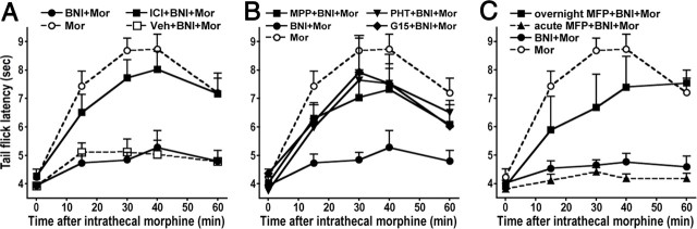
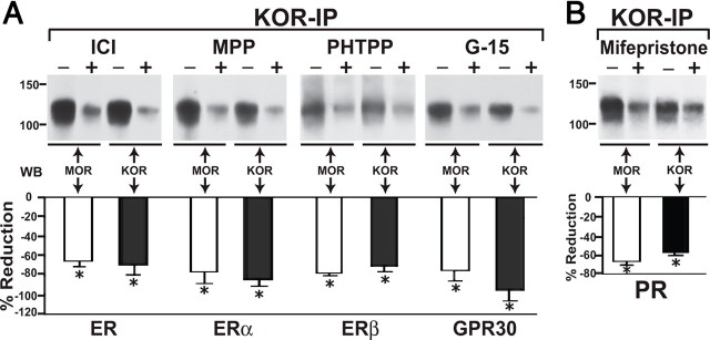
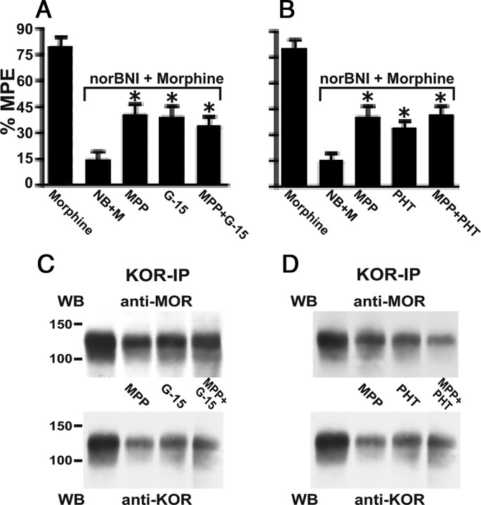
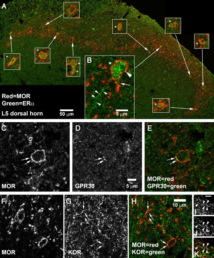
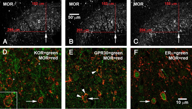
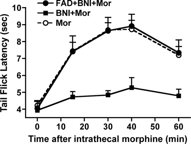
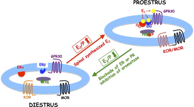
We previously demonstrated that the spinal cord κ-opioid receptor (KOR) and μ-opioid receptor (MOR) form heterodimers (KOR/MOR). KOR/MOR formation and the associated KOR dependency of spinal morphine antinociception are most robust during proestrus. Using Sprague Dawley rats, we now demonstrate that (1) spinal synthesis of estrogen is critical to these processes, and (2) blockade of either estrogen receptor (ER) α-, β-, or G-protein-coupled ER1 or progesterone receptor (PR) substantially reduces KOR/MOR and eliminates mediation by KOR of spinal morphine antinociception. Effects of blocking ERs were manifest within 15 min, whereas those of PR blockade were manifest after 18 h, indicating the requirement for rapid signaling by estrogen and transcriptional effects of progesterone. Individual or combined blockade of ERs produced the same magnitude of effect, suggesting that they work in tandem as part of a macromolecular complex to regulate KOR/MOR formation. Consistent with this inference, we found that KOR and MOR were coexpressed with ERα and G-protein-coupled ER1 in the spinal dorsal horn. Reduction of KOR/MOR by ER or PR blockade or spinal aromatase inhibition shifts spinal morphine antinociception from KOR dependent to KOR independent. This indicates a sex steroid-dependent plasticity of spinal KOR functionality, which could explain the greater analgesic potency of KOR agonists in women versus men. We suggest that KOR/MOR is a molecular switch that shifts the function of KOR and thereby endogenous dynorphin from pronociceptive to antinociceptive. KOR/MOR could thus serve as a novel molecular target for pain management in women.


Figures

Figure 1.
Figure 1.
The KOR-dependent spinal morphine antinociception in proestrus rats requires rapid ER signaling as well as transcriptional effects of PR. ER antagonists were administered together with morphine (5 μg) to the intrathecal space of proestrus rats that had been pretreated with intrathecal nor-BNI (BNI; 26 nmol) overnight. TFL was determined at various intervals thereafter. Effects of ER blockade could be observed as early as 15 min after antagonist administration. Data are shown as mean ± SEM. A, Intrathecal application of ICI 182,780 (ICI; ERα and ERβ antagonist) concomitant with morphine (MOR) abrogated the inhibitory effect of nor-BNI on morphine antinociception. B, Intrathecal application of MPP (ERα blocker), PHTPP (PHT; ERβ blocker), or G-15 (GPR30 blocker) concomitant with morphine also restored normative analgesic responsiveness to morphine, despite previous overnight treatment with nor-BNI. C, The overnight intrathecal pretreatment with mifepristone (MFP, PR antagonist) and nor-BNI negated the inhibition of spinal morphine antinociception that resulted from KOR blockade. The dose of ER and PR antagonists was 10 nmol. n = 11, 9, 7, 5, 5, 4 for ICI 182,780, MPP, PHTPP, G-15, MFP (overnight), and MFP (acute), respectively. Vehicles (Veh) used to administer the antagonists did not alter the effect of nor-BNI on spinal morphine antinociception. None of the ER or PR blockers themselves altered spinal morphine antinociception.
Figure 2.
Figure 2.
ER or PR blockade reduces KOR/MOR formation. A, Top, Immunoprecipitates obtained using anti-KOR antibodies from spinal cord of proestrus rats 30 min after intrathecal treatment with either vehicle or ICI 182,780 (ICI), MPP, PHTPP, or G-15 (10 nmol each) were Western blotted using anti-MOR antibodies and anti-KOR antibodies. An N-terminally directed anti-KOR antibody was used for IP, whereas the anti-KOR antibody used for Western blot analysis was generated against amino acids 262–275. The ≈120 kDa KOR/MOR signal was always detected by anti-MOR and anti-KOR antibodies. Blockade of ERα, ERβ, or GPR30 markedly reduced (∼70–80%; p < 0.05) spinal cord levels of KOR/MOR (n = 3–5). B, Immunoprecipitates obtained using anti-KOR antibodies from spinal cord of proestrus rats that had been pretreated overnight with intrathecal vehicle or mifepristone (10 nmol) were Western blotted (WB) using anti-MOR or anti-KOR antibodies. A striking reduction (≈62%) of the KOR/MOR Western blot signal was observed in mifepristone-pretreated rats versus no treatment using either antibody (n = 3). Bar graphs below Westerns blots in A and B shows percentage reduction (mean ± SEM) of KOR/MOR expression produced by blocking individual ERs or PR ascertained by Western blot analyses using anti-MOR antibodies (open bars) or anti-KOR antibodies (filled bars). In accordance with the behavioral data shown in Figure 1, rapid signaling of membrane ERs but transcriptional effects of PR signaling are both essential for elevated expression of KOR/MOR during proestrus. Notably, ER and PR antagonists produced similar reductions in KOR/MOR expression. *p < 0.05 for KOR/MOR expression in antagonist-treated versus untreated spinal cord that was analyzed in parallel.
Figure 3.
Figure 3.
Concurrent but not additive activities of ERα, ERβ, and GPR30 are required for the female-specific KOR-dependent spinal morphine antinociception and elevated spinal levels of heterodimeric KOR/MOR during proestrus. A, B, Intrathecal morphine (5 μg) was administered together with 1 pmol of MPP, 10 pmol of G-15, 15 pmol of PHTPP (PHT) or combinations thereof (MPP + G-15; MPP + PHTPP) to proestrus rats that had been pretreated overnight with intrathecal nor-BNI (26 nmol). TFL was determined 30 min after morphine treatment. Data show peak effect of drugs expressed as %MPE. C, D, Membranes obtained from spinal cord of proestrus rats that had been intrathecally treated for 30 min with vehicle (DMSO), one of two ER-type-selective blockers, or concomitantly with both were immunoprecipitated using anti-KOR antibodies. Immunoprecipitates were processed and Western blotted (WB) for KOR/MOR in parallel using anti-MOR and anti-KOR antibodies. Individual blockade of ERα, ERβ, or GPR30 partially restored spinal morphine antinociception despite nor-BNI pretreatment and partially reduced the levels of heterodimeric KOR/MOR. However, effects of MPP + G-15 or MPP + PHTPP on both measures were not significantly different from that which resulted from their individual application. *p < 0.05 for comparison between antinociception resulting from morphine + nor-BNI versus morphine + nor-BNI + ER type-selective antagonists. NB, nor-BNI; M, morphine.
Figure 4.
Figure 4.
Coexpression of MOR-IR with ERα, GPR30, or KOR in the superficial dorsal horn. A, B, MOR + ERα. A, A montage of the entire superficial dorsal horn of a single section of L5 spinal cord. Insets show 3× higher-magnification views of the cells pointed out by the arrows; cells labeled by ERα are marked with an asterisk (*). ERα-IR was found in 9 of the 10 MOR-IR cells. B, High-magnification view of a cell in the central-lateral superficial dorsal horn. ERα-IR was not restricted to the cell nucleus but also appeared to be in the plasma membrane (large arrowhead) and to extend into proximal dendrites (arrow). ERα-IR was also present in fibrous processes (small arrowheads). The 50 μm scale bar in A applies only to the large montage; magnification of insets is 3× higher. The 5 μm scale bar in B applies only to B. C–E, Expression of GPR30 by an MOR-IR cell in L6 superficial dorsal horn. Arrows mark MOR–GPR30 double labeling. GPR30-IR can also be seen within the cell cytoplasm. F–K, Coexpression of MOR-IR and KOR-IR. F–H, MOR–KOR coexpression in somata. Arrows mark KOR-IR structures visible in MOR-IR neurons in L5 superficial dorsal horn. Double labeling frequently appeared to be in or near the plasma membrane, but sometimes was also seen in the cytoplasm. I–K, MOR–KOR coexpression in fibrous processes. Arrowheads mark a fiber in L5 superficial dorsal horn that was double labeled for MOR and KOR. Scale bar (in I): I–K, is 2 μm.
Figure 5.
Figure 5.
MOR, GPR30, ERα, and KOR expressed in a single dorsal horn neuron. Confocal microscopic images are taken from three serially adjacent 5 μm cryostat sections. A–C, Lower-magnification images that provide overviews of the region. Arrows point to a single cell visible in all three sections expressing all four receptors. Dotted boxes outline the regions shown in D–F. D–F, Higher-magnification images that show double labeling for MOR and KOR (D), MOR and GPR30 (E), or MOR and ERα (F). Again, arrows point to a single cell that expresses all four receptors. D, Asterisks mark other MOR-IR cells that also express KOR-IR; inset shows a higher-magnification view of the cell marked by the arrow. Note green KOR-IR inside the MOR-IR cell. E, Arrowheads mark GPR30-IR in or near plasma membranes of MOR-IR neurons. (GPR30-IR is also visible inside the cytoplasm.) Note GPR30-IR in cell marked by arrow. F, Note ERα-IR in the MOR-IR cell marked by the arrow. Most MOR-IR neurons also expressed ERα (asterisks).
Figure 6.
Figure 6.
Inhibition of spinal cord aromatase eliminated the female-specific KOR-dependent spinal morphine (MOR) antinociception during proestrus. Fadrozole (FAD; 2.5 nmol) was administered to the intrathecal space of proestrus rats that had been pretreated overnight with nor-BNI (BNI; 26 nmol). One hour later, morphine (5 μg) was given intrathecally. TFL determination commenced 30 min thereafter. Intrathecal fadrozole significantly restored the nor-BNI blocked morphine antinociception.
Figure 7.
Figure 7.
Schematic representation of the modulation of KOR/MOR formation by E2 and P4. MOR, KOR, ERα, and GPR30 are coexpressed in neurons of the spinal dorsal horn. Biochemical and behavioral experiments suggest that ERs work in a cooperative manner as part of a macromolecular complex to increase KOR/MOR expression. We hypothesize that E2 (spinally synthesized and ovarian derived) triggers the formation of a signaling complex that contains multiple ERs, which via as of yet unknown mechanism(s) enhances heterodimerization of KOR and MOR. Transcriptional effects of P4 are essential either for the formation of the ER signaling complex and/or the heterodimerization of KOR with MOR.

References

    1. Albanito L, Madeo A, Lappano R, Vivacqua A, Rago V, Carpino A, Oprea TI, Prossnitz ER, Musti AM, Andò S, Maggiolini M. G protein-coupled receptor 30 (GPR30) mediates gene expression changes and growth response to 17beta-estradiol and selective GPR30 ligand G-1 in ovarian cancer cells. Cancer Res. 2007;67:1859–1866. - PubMed
    1. Arvidsson U, Riedl M, Chakrabarti S, Lee JH, Nakano AH, Dado RJ, Loh HH, Law PY, Wessendorf MW, Elde R. Distribution and targeting of a μ-opioid receptor (MOR1) in brain and spinal cord. J Neurosci. 1995a;15:3328–3341. - PMC - PubMed
    1. Arvidsson U, Riedl M, Chakrabarti S, Vulchanova L, Lee JH, Nakano AH, Lin X, Loh HH, Law PY, Wessendorf MW, Elde R. The k-opioid receptor (KOR1) is primarily postsynaptic: combined immunohistochemical localization of the receptor and endogenous opioids. Proc Natl Acad Sci U S A. 1995b;92:5062–5066. - PMC - PubMed
    1. Balthazart J, Baillien M, Ball GF. Rapid and reversible inhibition of brain aromatase activity. J Neuroendocrinol. 2001a;13:63–73. - PubMed
    1. Balthazart J, Baillien M, Ball GF. Phosphorylation processes mediate rapid changes of brain aromatase activity. J Steroid Biochem Mol Biol. 2001b;79:261–277. - PubMed

Publication types

MeSH terms