Review
. 2011 Aug 18;12(9):615-27.
doi: 10.1038/nrg3030.

Dynamic interactions between transposable elements and their hosts

Affiliations
Review

Dynamic interactions between transposable elements and their hosts

Henry L Levin et al. Nat Rev Genet. .

Abstract

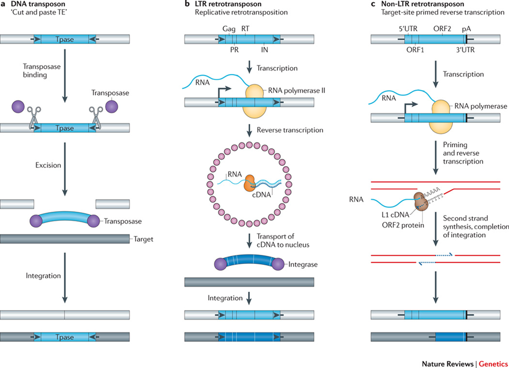
Transposable elements (TEs) have a unique ability to mobilize to new genomic locations, and the major advance of second-generation DNA sequencing has provided insights into the dynamic relationship between TEs and their hosts. It now is clear that TEs have adopted diverse strategies - such as specific integration sites or patterns of activity - to thrive in host environments that are replete with mechanisms, such as small RNAs or epigenetic marks, that combat TE amplification. Emerging evidence suggests that TE mobilization might sometimes benefit host genomes by enhancing genetic diversity, although TEs are also implicated in diseases such as cancer. Here, we discuss recent findings about how, where and when TEs insert in diverse organisms.


Figures

Figure 1
Figure 1. The diverse mechanisms of transposon mobilization
A. DNA transposons: Many DNA transposons are flanked by terminal inverted repeats (TIRs, black arrows), encode a transposase (TPase, purple circles), and mobilize by a “cut and paste” mechanism (scissors). Transposase binds at or near the TIRs, excises the transposon from its existing genomic location (light gray bar), and pastes it into a new genomic location (dark gray bar). The cleavages of the two strands at the target site are staggered, resulting in a target site duplication (TSD), typically 4 to 8 bp as specified by the TPase (Short black lines flanking the TE). B & C. Retrotransposons: Retrotransposons mobilize by a replicative mechanism that requires the reverse transcription of an RNA intermediate. B. LTR-retrotransposons contain two long terminal repeats (LTRs, black arrows) and encode Gag, protease (PR), reverse transcriptase (RT), and integrase (IN) activities critical for retrotransposition. The 5’ LTR contains a promoter that is recognized by the host RNA polymerase II and produces the mRNA of the TE (start of transcription indicated by a black vertical line attached to a right facing arrow). In step #1 of the reaction, Gag (small pink circles) assembles into virus like particles containing TE mRNA, RT, and IN. The RT copies the TE mRNA into a full-length double stranded DNA (wide blue arc). In step #2 of the reaction, IN (purple circles) inserts the DNA into the new target site. Similar to the TPases of DNA transposons, INs create staggered cuts at the target sites that result in TSDs. C. Non-LTR retrotransposons (right) lack LTRs and encode either one or two open reading frames. The transcription of non-LTR retrotransposons (indicate arrow as in panel b) also leads to the production of a full-length mRNA (blue wavy line). However, these elements mobilize by target-site primed reverse transcription (TPRT). An element-encoded endonuclease generates a single-strand “nick” in genomic DNA, liberating a 3’OH that is used to prime reverse transcription of the RNA. The proteins encoded by autonomous non-LTR retrotransposons also can mobilize non-autonomous retrotransposon RNAs, as well as other cellular RNAs (see text). The TPRT mechanism of an L1 element is depicted in the figure; the new L1 retrotransposition event is 5’ truncated and is retrotransposition-defective (bottom dark gray rectangle with blue line). Some non-LTR retrotransposons lack a poly A tails at their 3’ ends. The integration of non-LTR retrotransposons can lead to target-site duplications (TSDs) and small deletions at the target site in genomic DNA. For example, L1s are generally flanked by 7–20 bp TSDs.
Figure 2
Figure 2. Mechanisms that position integration
A. The Ty3 targeting mechanism: Integration of Ty3 occurs one or two bp upstream of tRNA genes. This pattern requires Brf1 and TBP, components of TFIIIB that recruit integrase (IN, gray oval) to the target site. B. The Ty1 targeting mechanism: The factor Bdp1 is a component of TFIIIB (green circles) that is required to recruit the chromatin remodeling complex Isw2 (light blue semi-circle). Integration of Ty1 occurs with an 80 bp periodicity in a 700 bp window upstream of tRNA genes. The periodicity requires both Bdp1 and Isw2. C. The mechanism of Tf1 targeting: Tf1 integrates into promoters transcribed by RNA polymerase II. Transcription factors (Act), such as Atf1p, bind to the promoter and recruit IN to the insertion sites. D. The mechanism of Ty5 integration: In the absence of a stress condition, the phosphorylation of the IN targeting domain (blue knob on TD) directs integration to heterochromatin by binding a structural component of the heterochromatin, Sir4 (green ovals). Orange cylinders indicated condensed nucleosomes; black lines represent DNA. When cells are exposed to environmental stress the IN is dephosphorylated and integration occurs in gene rich regions.
Figure 3
Figure 3. The degradation of transposon mRNA by RNAi
A. The siRNA pathway: Double-stranded “trigger” RNAs (hairpin), derived from the inverted terminal repeats of a DNA transposon in the case illustrated, are processed and then cleaved into 21 to 24 nt siRNAs by the dicer family of proteins (light green amorphous shape). A single-strand siRNA (short red line) complementary to the transposon mRNA is selectively incorporated into the Argonaute containing RISC complex (blue amorphous shape with short red line). The siRNA directs RISC to complementary sequences in transposon mRNA (long red line), leading to its post-transcriptional destruction. The figure was drawn based on concepts presented in the following reviews, , . B. The piRNA pathway: A primary piRNA transcript (wavy blue line) generated from a piRNA cluster (blue rectangle) that contains sequences derived from TEs (darker blue rectangle inset) is processed into mature 24–35 nt piRNAs (small blue line). Binding of the mature piRNA by the Piwi/Aubergine protein (color?…make different from Ago3 and panel A) allows it to be directed to complementary sequences in TE mRNA (red line). Endonucleolytic cleavage (scissor) 10 nt from the 5’ end of the small RNA and 3’ processing liberates a secondary sense strand transposon piRNA (red line), which associates with the Argonaute 3 protein (color?…make different from Aubergine and panel A). The binding of this complex to complementary sequences in the original precursor piRNA, followed by endonucleolytic cleavage and 3’ processing liberates, generates a secondary antisense piRNA that can be directed to TE mRNA. This reiterative cycle (e.g., a “ping-pong” cycle) can lead to the destruction of transposon mRNA in the germ line. The piRNA model was redrawn based on concepts and models presented in the following papers, , , and for the example illustrated is from Drosophila; however, a similar pathway likely operates in mammalian cells (see text for details).
Figure 4
Figure 4. Timing of transposition
TE mobility in cells giving rise to gametes, as well as TE mobility post-fertilization during early development, can lead to germline TE integration events. Embryonic TE mobility in cells that do not contribute to the germline, or at later times in development can, in principle, lead to somatic TE integration events. The overlapping brackets signify that some TE insertions in early development can contribute to the germline, whereas others may not. Endogenous L1 retrotransposition events can occur in certain tumors, and experiments using engineered human L1s suggest that L1 retrotransposition also may occur during mammalian neurogenesis. Examples of the developmental timing of TE integration events are described in the main text.

References

    1. McClintock B. The origin and behavior of mutable loci in maize. Proc Natl Acad Sci U S A. 1950;36:344–355. - PMC - PubMed
    1. Levis RW, Ganesan R, Houtchens K, Tolar LA, Sheen FM. Transposons in place of telomeric repeats at a Drosophila telomere. Cell. 1993;75:1083–1093. - PubMed
    1. Agrawal A, Eastman QM, Schatz DG. Transposition mediated by RAG1 and RAG2 and its implications for the evolution of the immune system. Nature. 1998;394:744–751. - PubMed
    1. Feschotte C. Transposable elements and the evolution of regulatory networks. Nat Rev Genet. 2008;9:397–405. - PMC - PubMed
    1. Beck CR, Garcia-Perez JL, Badge RM, Moran JV. LINE-1 Elements in Structural Variation and Disease. Annu Rev Genomics Hum Genet. 2011;12 in press. - PMC - PubMed

Publication types