

Retinal nerve fiber hypertrophy in ataxia of Charlevoix-Saguenay patients
- PMID: 21850161
- PMCID: PMC3144729
Retinal nerve fiber hypertrophy in ataxia of Charlevoix-Saguenay patients
Abstract
Purpose: To present full ophthalmologic examination and retinal nerve fiber layer (RNFL) photographs of autosomal recessive spastic ataxia of Charlevoix-Saguenay (ARSACS) patients showing significant increases in RNFL thickness compared to healthy subjects, but without myelinated retinal fibers.
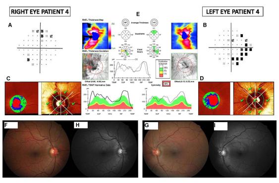
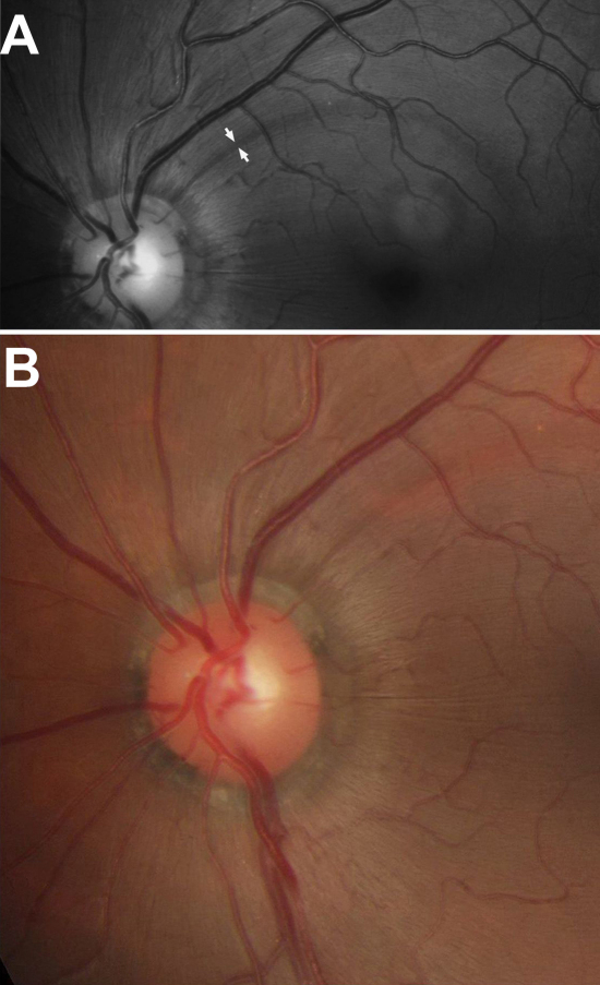
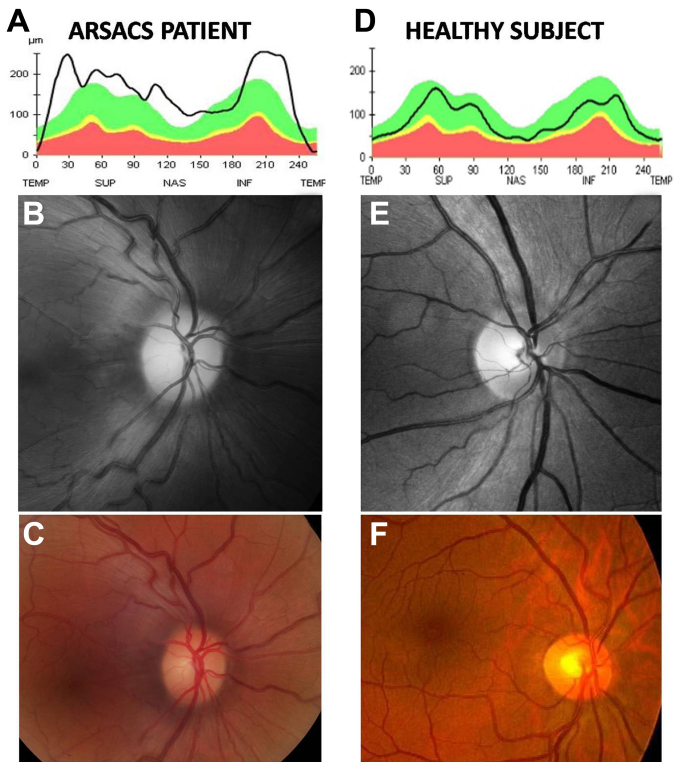
Methods: The study design was observational case series. Ten eyes of five patients with molecular confirmation of ARSACS underwent a full ophthalmologic examination that included clinical history, visual acuity, biomicroscopy of the anterior segment, gonioscopy, Goldmann applanation tonometry, central corneal ultrasonic pachymetry, ophthalmoscopy of the posterior segment, standard automatic perimetry (Humphrey field), simultaneous stereophotographs of the optic disc after mydriasis, a series of five red-free digital fundus photographs for RNFL evaluation, topographic analysis of the optic disc using the Heidelberg retina tomography, and measurement of peripapillary RNFL thickness with Cirrus optical coherence tomography.
Results: All patients showed abnormal visual fields, normal optic discs with a mild to strikingly increased visibility of RNFL in color stereophotographs, normal Heidelberg tomography, and moderate to markedly increased RNFL thickness in Cirrus tomography (average thickness ranging from 119 μm to 220 μm).
Conclusions: We found evidence of RNFL hypertrophy in ARSACS patients that may have been interpreted as hypermyelinated retinal fibers in previous reports. A revision of ARSACS diagnostic criteria, particularly with regard to retinal alterations, is necessary.
Figures
References
-
- Engert JC, Bérubé P, Mercier J, Doré C, Lepage P, Ge B, Bouchard JP, Mathieu J, Melançon SB, Schalling M, Lander ES, Morgan K, Hudson TJ, Richter A. ARSACS, a spastic ataxia common in northeastern Québec, is caused by mutations in a new gene encoding an 11.5-kb ORF. Nat Genet. 2000;24:120–5. - PubMed
-
- De Braekeleer M, Giasson F, Mathieu J, Roy M, Bouchard JP, Morgan K. Genetic epidemiology of autosomal recessive spastic ataxia of Charlevoix-Saguenay in northeastern Quebec. Genet Epidemiol. 1993;10:17–25. - PubMed
-
- Dupré N, Bouchard JP, Brais B, Rouleau GA. Hereditary ataxia, spastic paraparesis and neuropathy in the French-Canadian population. Can J Neurol Sci. 2006;33:149–57. - PubMed
-
- Gerwig M, Krüger S, Kreuz FR, Kreis S, Gizewski ER, Timmann D. ARSACS outside Quebec Characteristic MRI and funduscopic findings help diagnose. Neurology. 2010;75:2133. - PubMed
-
- El Euch-Fayache G, Lalani I, Amouri R, Turki I, Ouahchi K, Hung WY, Belal S, Siddique T, Hentati F. Phenotypic features and genetic findings in sacsin-related autosomal recessive ataxia in Tunisia. Arch Neurol. 2003;60:982–8. - PubMed
MeSH terms
Supplementary concepts
LinkOut - more resources
Full Text Sources
Medical